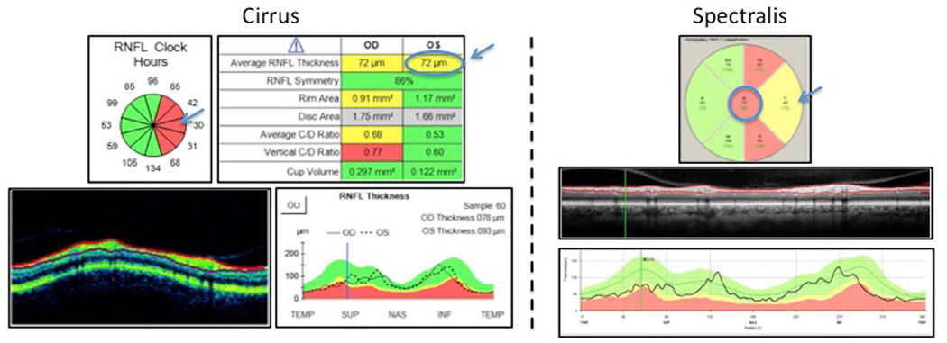
Figure 1.
Case 1. Global RNFL Classification Disagreement between Cirrus (borderline) and Spectralis (outside normal limits) in Glaucoma Suspects
Left: Cirrus scan showing global retinal nerve fiber layer classification as yellow, borderline (top right arrow), while most temporal clock hours are red, outside normal limits (left arrow). Right: Spectralis showing global retinal nerve fiber layer classification as red, outside normal limits (top arrow), while most temporal superior and temporal inferior sectors are red, outside normal limits (left arrow). Although global classification differs, both instruments identify temporal sectors (Spectralis) and clock hours (Cirrus) as outside normal limits.

