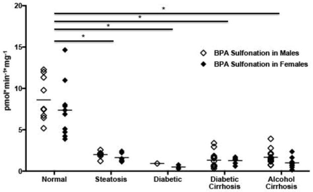
Figure 1. Bisphenol A (BPA) sulfonation is decreased in cytosolic fractions isolated from diseased human livers.
Each data point represents a single tissue (average of determinations) categorized by gender and disease type. (n=20 for nonfatty, n=13 for steatosis, n=4 for diabetes, n=21 diabetic cirrhosis, and n=21 for alcohol cirrhosis). Females are displayed as black diamonds (◆), males as white diamonds (□). BPA sulfonation was determined by incubating 4 μM bisphenol A with human liver cytosols for 30 min in the presence of the 35S-labeled cofactor 3′-Phosphoadenosine-5′-phosphosulfate (PAPS). The reaction rate was calculated as product formation (pmol)/reaction time (min) / amount of enzyme used (mg). Asterisks (*) represent statistically significant differences between non-fatty and diseased human livers.

