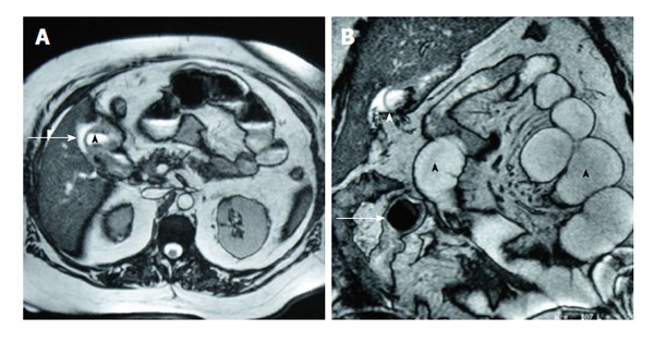
Figure 4.

Magnetic resonance cholangiopancreatography findings in a patient with gallstone ileus. A: On T2-MRI, a hyperintense image is identified in the gallbladder bed (arrow), with communication with the duodenal second portion (arrowhead), suggestive of a cholecystoduodenal fistula; B: MRI coronal reconstruction showed dilated small bowel loops with endoluminal air (black arrowheads) and a signal-void round-shaped image, suggestive of a gallstone (arrow). Gallbladder communication with duodenum is observed (white arrowhead). MRI: Magnetic resonance imaging.
