
Figure 2.
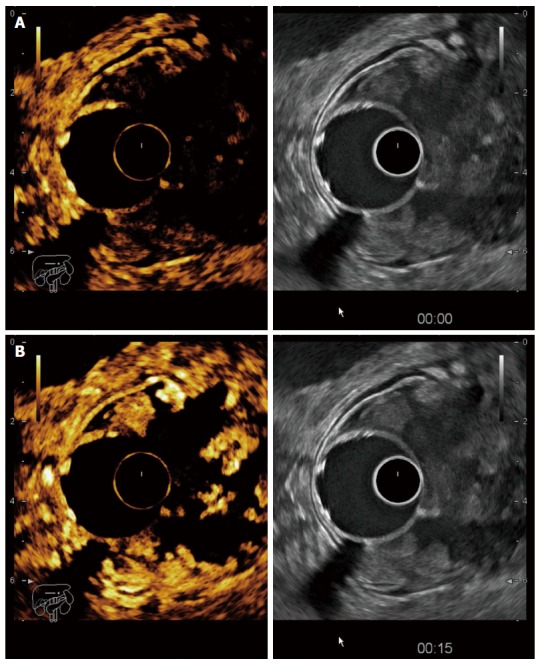
Contrast-enhanced endoscopic ultrasonography in a T3 tumour of the recto-sigmoid junction. A: Before contrast arrival (left side contrast harmonic imaging mode, right side B mode); B: Maximal enhancement of the tumour 15 s after contrast injection with hyperenhanced areas alternating with avascular (necrotic) areas.