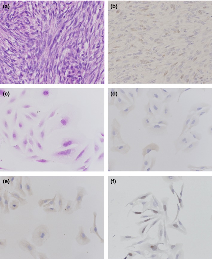
Figure 3.

Hematoxylin–eosin stain (a) and immunohistochemical stain for FOXM1 (b) of the original tumor specimen of TC616 soft tissue leiomyosarcoma cells. Light microscopic findings of TC616 cells in vitro. The tumor cells were spindle, round, or polygonal in shape with oval nuclei and extension of slender cytoplasmic processes (c). Most TC616 cells showed immunopositive reactions for α‐smooth muscle actin (d), muscle‐specific actin (e), and FOXM1 (f) antibodies.
