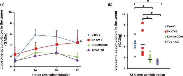
Figure 3.

Ex vivo accumulation of 111In‐encapsulated liposomes at several time points among four human ovarian cancer xenografted tumors. (a) Ex vivo analysis at each time point revealed significantly higher accumulation levels in Caov‐3 and SK‐OV‐3 tumors than those in KURAMOCHI and TOV‐112D tumors at 72 h after injection. Six to 11 mice were used for analysis in each group at each time point. (b) Accumulation of 111In‐encapsulated liposomes in an individual mouse at 72 h after injection. Most notably, among all four tumor types analyzed, only SK‐OV‐3 displayed a wide range of variation. *P < 0.05. %AD/g, % of administered dose/gram of organ.
