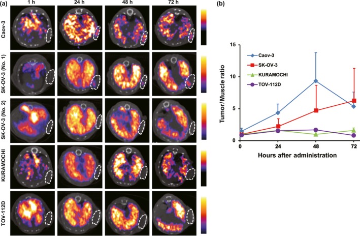
Figure 4.

Single‐photon emission computed tomography (SPECT)/CT imaging of 111In‐encapsulated liposomes in human ovarian cancer mouse xenograft models. (a) Representative axial SPECT/CT images of mouse xenograft models (n = 3–5). White circle indicates tumor region based on superimposed CT image. High accumulation levels were observed in Caov‐3, followed by SK‐OV‐3, tumors between 24 and 72 h. We observed interindividual variation in SK‐OV‐3 xenograft mice from 48 h with clear depiction of tumor (No. 1) or none (No. 2). (b) Tumor/background ratio from SPECT image data analysis.
