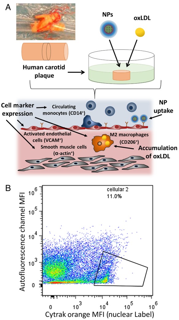
Figure 2.

(A) Plaque specimens were excised from human carotid arteries, sectioned into uniform 2–5 mm cylindrical sections, and treated with fluorescent AM NPs (1 × 10⁻⁵ M) and 5 µg/mL DiO oxLDL. Cell subsets within the plaque were evaluated for NP and oxLDL uptake, including CD206+ (surface protein of M2 phenotype), VCAM+ (surface protein of inflamed endothelial cells), CD14+ (surface protein of monocytes and macrophages), and α-actin+ (surface and intracellular protein of smooth muscle cells). (B) To ensure proper evaluation via flow cytometry of the cellular population within the plaque, Cytrak orange was used as a nuclear label, and only populations positive for this marker were used in the data analysis.
