
Figure 2.
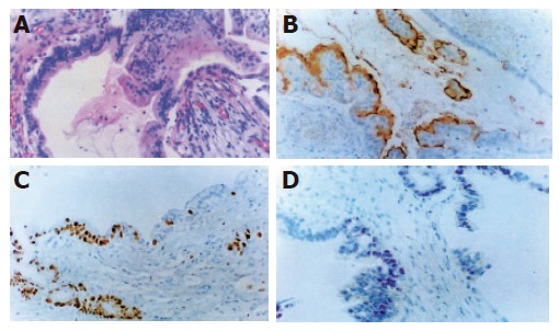
H&E and immunohistochemical staining of dysplastic tissues of the pancreatic duct. A: The nuclear enlargement, increased nuclear-to-cytoplasmic ratio, loss of nuclear polarity, pleomorphism, and nuclear overcrowding are seen. Several mitotic figures including atypical form are present. H&E stain, ×100; B: CEA monoclonal antibody shows positive reaction in the cytoplasmic membranes of dysplastic cells. DAB and hematoxylin counter stain, ×100; C: Ki-67 monoclonal antibody shows focally increased proliferating nuclei of dysplastic cells. DAB and hematoxylin counter stain, ×100; D: p53 monoclonal antibody shows focally positive reaction in the nuclei of dysplastic cells. AEC and hematoxylin counter stain, ×100.