
Figure 2.
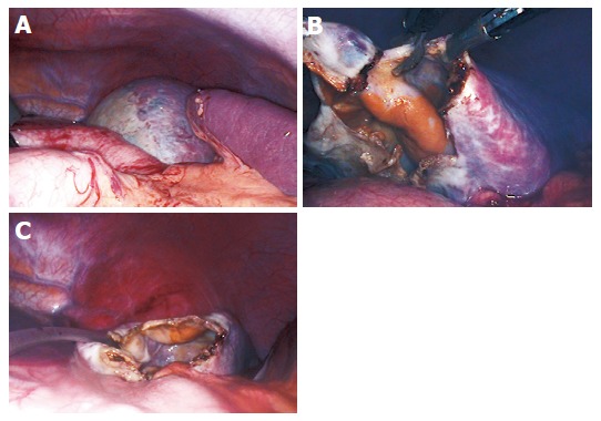
Laparoscopic view of the cyst. Most parts of the cyst are covered with a thin layer of splenic tissue; only a small portion in the upper pole of the spleen displays a “white roof” (A). The cyst was punctured and evacuated and a 3 cm×3 cm portion of the cyst was excised using the monopolar scissor (B). A drainage tube was inserted in the remaining cavity (C).