
Figure 2.
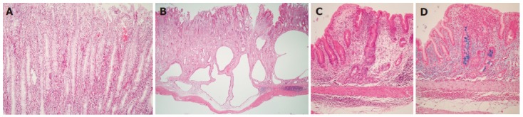
Microscopic views of the gastric body of Mongolian gerbils at 18 mo after H pylori inoculation. A: Severe infiltration of polymorphonuclear and mononuclear cells were seen in the lamina propria. (HE stain, x100); B: Some glands have extended into the submucosa but not into the proper muscularis layer. Severe infiltration of mononuclear cells in the submucosa (HE stain, x10); C: Intestinal metaplasia is seen scattering in gastric mucosa (HE stain, x10); D: Intestinal metaplasia (Alcian blue stain (pH 2.5); original magnification, x10).