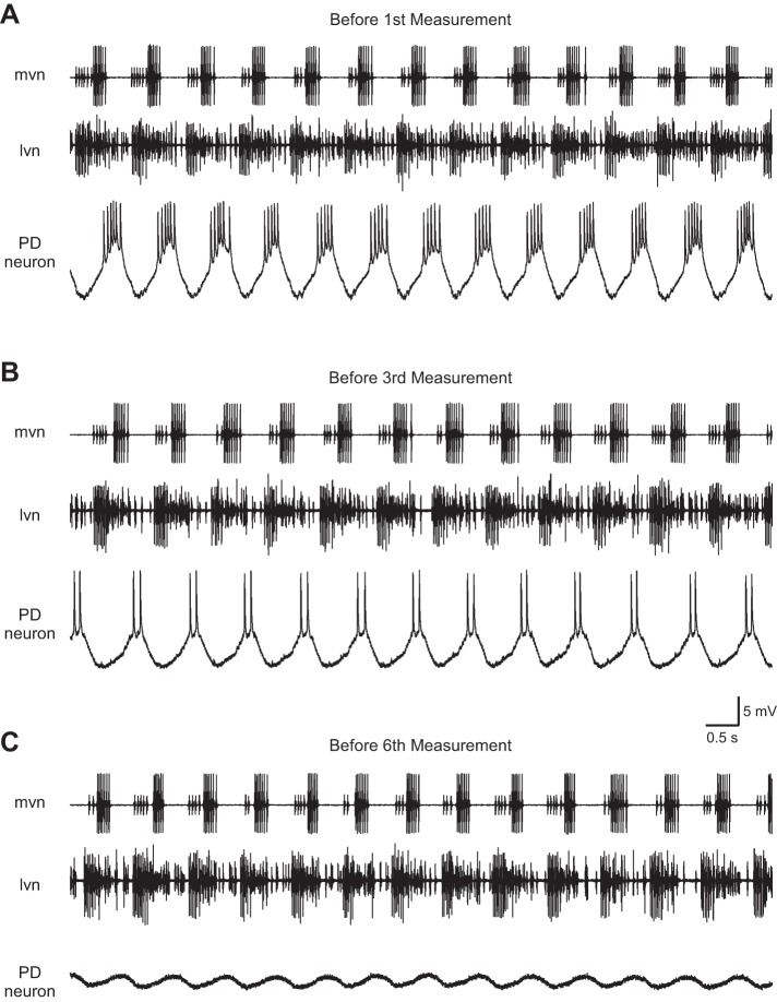
Fig. 14.
Raw data showing effect on PD neuron activity of recording with an unbeveled 6.5 M KAc electrode. Before the 1st measurement stimulation, the neuron fired 5–6 spikes/cycle (A); before the 3rd, only 2 spikes/cycle (B); and before the 6th, 0 spikes/cycle (C). Effect was not due to generalized cessation of pyloric network activity (note continued spiking activity on the extracellular traces). mvn, median ventricular nerve: large amplitude spikes are due to ventricular dilator neuron and small spikes, inferior cardiac neuron. lvn, lateral ventricular nerve: large spikes are due to lateral pyloric neurons and small spikes, pyloric and PD neuron activity (which cannot be well distinguished in this recording).

