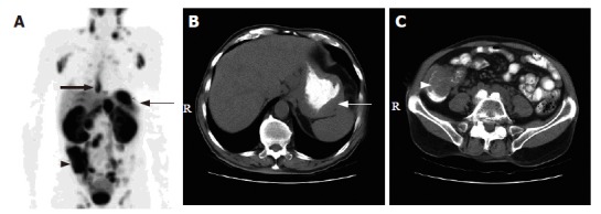
Figure 4.

A 78-year-old man with mantle cell lymphoma. A: 18F-FDG PET scan revealed multiple hypermetabolic foci involving the lower esophagus (thick arrow), gastric fundus (thin arrow), cecum and terminal ileum (arrow head); B and C: The corresponding CT scans of the abdomen showed wall thickening at the gastric fundus (white arrow) and the cecum and terminal ileum (white arrow head)
