Abstract
AIM: To test whether antioxidant treatment could prevent the progression of Barrett’s esophagus to adenocarcinoma.
METHODS: In a rat model of gastroduodenoesophageal reflux by esophagojejunal anastomosis with gastric preservation, groups of 6-10 rats were randomized to receive treatment with superoxide dismutase (SOD) or vehicle and followed up for 4 mo. Rat’s esophagus was assessed by histological analysis, superoxide anion and peroxinitrite generation, SOD levels and DNA oxidative damage.
RESULTS: All rats undergoing esophagojejunostomy developed extensive esophageal mucosal ulceration and inflammation by mo 4. The process was associated with a progressive presence of intestinal metaplasia beyond the anastomotic area (9% 1st mo and 50% 4th mo) (94% at the anastomotic level) and adenocarcinoma (11% 1st mo and 60% 4th mo). These changes were associated with superoxide anion and peroxinitrite mucosal generation, an early and significant increase of DNA oxidative damage and a significant decrease in SOD levels (P<0.05). Exogenous administration of SOD decreased mucosal superoxide levels, increased mucosal SOD levels and reduced the risk of developing intestinal metaplasia beyond the anastomotic area (odds ratio = 0.326; 95%CI: 0.108-0.981; P = 0.046), and esophageal adenocarcinoma (odds ratio = 0.243; 95%CI: 0.073-0.804; P = 0.021).
CONCLUSION: Superoxide dismutase prevents the progression of esophagitis to Barrett’s esophagus and adenocarcinoma in this rat model of gastrointestinal reflux, supporting a role of antioxidants in the chemoprevention of esophageal adenocarcinoma.
Keywords: Barrett’s esophagus, Esophageal adenocarcinoma, Free radicals, Oxidative damage, Superoxide dismutase
INTRODUCTION
The incidence of esophageal adenocarcinoma (EAC) has increased dramatically in Western countries in recent years[1]. Today, the existence of a cause-effect relationship between gastroesophageal reflux disease (GERD) and EAC is firmly accepted. In order to design preventive strategies that might stop/delay the malignant transformation of the esophageal epithelium under the impact of chronic gastroesophageal reflux, it is important to define the mechanisms and mediators of the inflammatory process driving the esophageal mucosa from chronic inflammation to adenocarcinoma. Recent studies have implicated reactive oxygen species (ROS) in the pathogenesis of GERD, Barrett’s esophagus (BE) and esophageal cancer. Increased levels of ROS have been found in esophageal tissues of patients with esophagitis and Barrett’s epithelium[2]. Furthermore, a positive correlation between the severity of esophagitis and ROS levels has been demonstrated, with the highest levels in patients with BE. At the same time, the free-radical scavenger superoxide dismutase (SOD) level is decreased in esophageal tissue of patients with gastroesophageal reflux, being lowest in patients with severe esophagitis and BE[3,4]. Studies performed in animal models of both acute and chronic reflux esophagitis have suggested a pathogenic role of ROS, especially superoxide anion, in the development of esophageal mucosal damage. Moreover, administration of SOD has been shown to either prevent or reduce mucosal damage[5-8]. However, it is not known whether SOD is effective in preventing the development of BE and EAC. In order to answer this question, we used an experimental model of gastroenteroesophageal reflux in rats that could lead to the development of EAC, to determine the temporal sequence of development of intestinal metaplasia (IM) and EAC and to assess the involvement of superoxide anion as a mechanism of damage. Finally, we tested whether administration of SOD was able to prevent the development of BE and/or EAC.
MATERIALS AND METHODS
Rat experimental model
All animal studies were carried out in the Service of Biomedicine and Biomaterials of the University of Zaragoza, officially inscribed as a “Research Establishment” for the adequate husbandry and use of all research animals under the “Good Laboratory Practices” norms and Spanish laws (RD 223/1988). The Ethics Committee approved all the procedures. Wistar rats weighing 250-300 g were studied. Rats underwent esophagojejunostomy with gastric preservation, allowing the gastroduodenal content to flow back into the esophagus. In a clean operating field, a midline abdominal incision was made and the liver was retracted to expose the stomach and intra-abdominal portion of the esophagus. Bilateral vagus nerves were preserved and the abdominal esophagus was transected proximal to the cardia and the distal cut end was closed with sutures. The esophagus was anastomosed end-to-side to an enterostomy performed in a jejunum loop distal to Treitz’s ligament with 12 interrupted stitches of all layers using 7/0 silk sutures. Sham operation consisted of laparotomy and blunt manipulation of abdominal contents. After the operation, rats were housed in hanging cages to prevent bed ingestion and fasted until d 3 with free access to drinking water. Buprenorphine was used as an analgesic.
Study design and drug administration
For the first purpose of the study, groups of animals (6-10 rats/group) were killed at 1 mo intervals up to 8 mo after esophagojejunostomy or sham operation. Since a high incidence of EAC and IM was found 4 mo after surgery, this follow-up was considered for the second part of the study, where the effect of SOD administration on the development of BE and EAC was tested. SOD (Orgotein-Ontosein®, Tedec-Zambaletti, Madrid, Spain) was administered at a dose of 3 mg/kg (10 000 U/kg) every 3 d subcutaneously. The SOD used was a liver bovine metalloprotein which has been shown to be highly effective in the treatment of secondary effects of radiotherapy on human head and neck tumors[9] and in reducing acid and pepsin-induced esophagitis in rabbits[5,6]. Previously, we have confirmed that the dose of SOD administered to rats increased blood levels of SOD activity for several hours, which returned to basal levels approximately 24 h after SOD administration. However, given the duration of the study and the need for long-term parenteral treatment, we decided to administer SOD to rats once in every 72 h, mimicking the dosage used in patients receiving radiotherapy. Groups of rats (6-10 rats/group) with esophagojejunostomy receiving SOD treatment were killed at 1 mo intervals. Control rats with esophagojejunostomy were administered vehicle following the same pattern as that of SOD treatment.
Rats were killed by CO2 inhalation. The esophagus was removed and opened longitudinally. More than half of the esophageal specimen was prepared for pathological study. The rest of the esophagus was cut into two slices: the lower and the upper half of the esophagus. For superoxide anion measurements, samples were processed immediately. For the rest of the determinations, samples were frozen immediately in liquid nitrogen and then kept at -70 °C until biochemical studies were performed.
Macroscopic and microscopic evaluation
The tissues were examined by two independent pathologists who were unaware of the experimental conditions. Changes of the squamous epithelium were classified into the following four categories. Reactive changes: characterized by the presence of basal cell hyperplasia, papillomatosis and hyperqueratosis with areas of inflammation and ulceration; Barrett’s esophagus: squamous epithelium was replaced with columnar-lined epithelium comprising occasional and incomplete differentiation of goblet cells, the length must be superior to 3 mm from the anastomosis site or intercalated with squamous epithelium and metaplasia of cells beyond the anastomosis or containing areas of dysplasia; Dysplasia: characterized by nuclear atypia, partial loss of mucosecretory function and cell polarity, and an increase in mitotic figures; Adenocarcinoma: glandular structures of epithelial cells with architectural dysplasia and cytologic atypia with stromal invasion and deep infiltration. In general, the two pathologists showed a high concordance (kappa coefficient >0.8). However, in case of disagreement, a third pathologist was required to resolve the diagnosis.
Measurement of superoxide anion, peroxynitrite, and SOD activity
The presence of superoxide anion was determined by chemiluminescence using lucigenin (N-methyl-acridinium nitrate) as previously described[10]. Results were expressed as counts per minute per milligram of fresh tissue (CPM/mg). Total SOD activity was measured according to a previously described method[10] based on the inhibition of nitro blue tetrazolium (NBT) mediated by superoxide anion. Results were expressed as units of SOD per gram of fresh tissue (U/g). The presence of peroxynitrites was indirectly measured by nitrotyrosine immunohistochemistry using a monoclonal anti-nitrotyrosine antibody (Chemicon, Temecula, CA, USA). Slides were examined microscopically and scored for relative staining intensity of nitrotyrosine: 0 (negative), 1+ (weak), 2+ (medium), 3+ (strong).
Nitrotyrosine immunoprecipitation
In order to purify the proteins containing tyrosine-nitrated residues, immunoprecipitation was performed using a monoclonal anti-nitrotyrosine antibody as previously described[11,12]. For this purpose, total protein was extracted from rat’s esophagus and protein concentrations were measured by Bradford assay (Bio-Rad, Hercules, CA, USA). Extracted proteins (500 µg) were precleared (3 h at room temperature) with 25 μL of protein G-agarose (Roche Applied Science, Mannheim, Germany). After a brief centrifugation, the supernatants were collected and incubated (2 h at room temperature) with 20 μL of anti-nitrotyrosine agarose conjugate (Alexis Biochemicals, Lausen, Switzerland). After the incubation, beads were collected, resuspended in 25 µL of 1× Laemmli sample buffer and boiled for 5 min. Then, the supernatants were immediately subjected to electrophoresis in 12% SDS polyacrylamide gels.
Western blot analysis
For analysis of Cu, Zn-SOD and Mn-SOD expression and detection of nitrated Mn-SOD, Western blot analysis was performed in both total protein extracts (60 μg of total protein) and immunoprecipitated fractions, respectively. After separation by SDS/PAGE, proteins were transferred electrophoretically to PDVF membranes, which were blocked (4 °C, overnight) with 5% non-fat dry milk in 50 mmol/L Tris-HCl, pH 7.4/150 mmol/L NaCl/0.05% Tween 20 (TBST). For detection of Mn-SOD, blots were incubated (1 h, room temperature) with a rabbit polyclonal anti-MnSOD antibody (1:1 000 dilution, Upstate, Charlottesville, VA, USA). For the detection of Cu, Zn-SOD, blots were incubated (1 h, room temperature) with a rabbit polyclonal anti-Cu, Zn-SOD antibody (1:1 000 dilution, Santa Cruz Biotechnology Inc., Santa Cruz, CA, USA). After incubation with primary antibodies, membranes were probed (1 h, room temperature) with 1:3 000 dilution of peroxidase-conjugated secondary antibody in TBS with 0.1% of blocking agent. Immunoreactive proteins were detected using enhanced chemiluminescence (ECL Western blotting analysis system, Amersham Biosciences, Buckinghamshire, UK).
Measurement of 8-hydroxy-2’-deoxyguanosine levels in esophagus
Genomic DNA was extracted from the lower half of the esophagus and digested with DNAse I, alkaline phosphatase, nuclease P1 and phosphodiesterases I and II (Roche Molecular Bio-Chemicals, Indianapolis, IN, USA), in order to obtain free nucleosides according to previously described methods[13]. The nucleoside samples were used for the determination of the amount of 8-hydroxy-2’-deoxyguanosine, which was measured using a competitive enzyme-linked immunosorbent assay kit (Bioxytech 8-OHdG-EIA kit, OXIS Health Products, Portland, USA). Thirty micrograms of digested DNA was added to each well. Each sample was performed in triplicate. Results were expressed as nanograms of 8-OHdG per milligram of total DNA.
Statistical analysis
Data management and statistical analysis were performed using the SPSS software v.10.1. Results from biochemical assays were expressed as mean±SE. Data were compared between groups by non-parametric tests (Kruskall-Wallis and Mann-Whitney). P<0.05 was considered statistically significant.
Covariance analysis was used for the analysis of the drug on the quantitative variables, introducing the number of months as covariant. Multiple comparisons were corrected by means of the Bonferroni method. Logistic models were adjusted for the qualitative variables, evaluating in turn the effect of the drug and the months passed.
RESULTS
Pathological findings
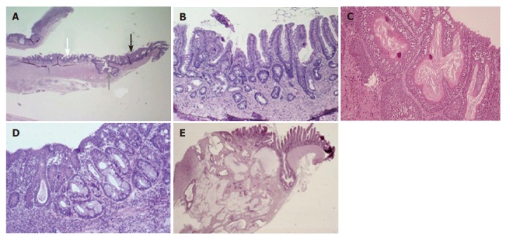
Macroscopically, all rats with esophagojejunostomy showed dilatation and thickening of middle and lower esophagus. The inner surface of the esophagus showed ulcerations, which were larger the closer they were to the distal esophagus. Macroscopically, no esophageal tumors were identified. Microscopically, in most animals, the upper and the middle parts of the esophagus showed squamous hyperplasia. Squamous epithelium also showed extensive defects, in the form of erosions in the upper third or ulcers in the proximity of the anastomosis. In all rats with esophagojejunostomy, columnar-lined metaplasia was found in the area of anastomosis, in continuity with the jejunal epithelium (Figures 1A and 1B) after 5 mo. A second type of metaplasia was found far above the anastomosis in some animals. This metaplasia consists of isolated or grouped goblet cells, which formed authentic mucus glands in some cases (Figures 1C and 1D). In all cases an important inflammatory infiltrate was present. Neoplastic transformation of the epithelium was always found near the anastomosis, surrounded by columnar epithelium. Histologically, all tumors were adenocarcinomas, most of them mucinous (70.37%) (Figure 1E).
Figure 1.

Pathological findings in the esophagus of rats with esophagojejunostomy. (A) Development of intestinal metaplasia in the area of anastomosis contiguous to the jejunal epithelium. Characteristic jejunal epithelial villi (white arrow) contrast with blunt villi of metaplastic areas (black arrow). The gray arrow shows the site of anastomosis (H&E staining, ×12.5); (B) A higher magnification of (A) shows the metaplastic epithelium in detail, with incomplete development of villi and crypts lined by absorptive cells with brush borders and goblet cells (H&E, ×100); (C) Intestinal metaplasia developed distant from the anastomosis, consisting of isolated goblet cells immersed within squamous epithelium (H&E, ×100); (D) Formation of mucus glands in squamous epithelium; (E) Development of mucinous adenocarcinoma from metaplasia in continuity, infiltrating the esophageal wall in depth. The site of anastomosis is shown by the gray arrow (H&E, ×12.5).
Incidence of lesions over time
Only rats that were killed in the protocol time were included in the study of the sequence esophagitis-BE-dysplasia-adenocarcinoma. The outcome of the rats included in different groups is shown in Table 1. Sham-operated rats did not show any esophageal change after 8 mo of follow-up. Inflammation, basal cell hyperplasia, hyperqueratosis and ulceration were present in mo 1. Columnar metaplasia was present in the first month, reaching an incidence of 100% in mo 2. The length of metaplasia increased progressively over time, with an average length of 1.61 mm in the first month to
Table 1.
Histological findings after surgery
| Mo | 1 (n = 11) (%) | 2 (n = 11) (%) | 3 (n = 8) (%) | 4 (n = 16) (%) | 5 (n = 11) (%) | 6 (n = 8) (%) | 7 (n = 11) (%) | 8 (n = 8) (%) |
| Reactive epithelium, ulceration, inflammation | 100 | 100 | 100 | 100 | 100 | 100 | 100 | 100 |
| Intestinal metaplasia in continuity to anastomosis | 66.6 | 100 | 100 | 93.75 | 100 | 100 | 100 | 100 |
| Intestinal metaplasia beyond the anastomosis | 9.09 | 70 | 62.5 | 50 | 63.63 | 75 | 40 | 70 |
| Dysplasia | 36.36 | 85.12 | 100 | 93.75 | 100 | 100 | 100 | 75 |
| Adenocarcinoma | 11.11 | 54.54 | 87.5 | 60 | 90.9 | 75 | 80 | 75 |
6.75 mm in mo 8. Metaplasia also showed a progressive differentiation over time. Metaplasia beyond the anastomosis was identified in the first month, but increased substantially after 2 mo. Dysplasia and adenocarcinoma rates increased dramatically in the second month.
Superoxide anion generation, SOD activity and nitrotyrosine immunohistochemistry
An early and significant increase in superoxide anion generation and a significant decrease in SOD activity were evident in the second month after esophagojejunostomy (Figures 2A and 3A). When grouping the animals according to the highest lesion found in the sequence BE-dysplasia-adenocarcinoma, all groups showed higher superoxide anion levels than control rats (P<0.05), reaching the highest level in those rats with BE (Figure 2B). In parallel to these changes, a significant decrease in SOD activity was observed in all groups of rats with mucosal lesions when compared to control rats (Figure 3B).
Figure 2.

Superoxide anion levels in distal esophagus at different time points (A) and in different types of lesion (B) after esophagojejunostomy. aP<0.05 vs control group (sham-operated rats) (n = 6-10).
Figure 3.

Superoxide dismutase levels in distal esophagus at different time points (A) and in different types of lesion (B) after surgery. aP<0.05 vs control group (sham-operated rats) (n = 6-10).
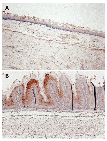
In sham-operated rats, nitrotyrosine staining was absent or weak in the esophagus (Figure 4A). In contrast, in rats with esophagojejunostomy, a positive nitrotyrosine staining was found in all rats (55% grade 2, 45% grade 1). Interestingly, the intensity of staining was stronger in squamous epithelium, while weaker positive staining was seen in columnar epithelium (Figure 4B).
Figure 4.

Representative nitrotyrosine immunohistochemical staining in the distal esophagus of rats without reflux (sham operation), showing weak and superficial staining (A) and stronger staining (B) in the cytoplasm of squamous epithelial cells of rats after 8 weeks of reflux (esophagojejunostomy).
SOD expression
The expression of MnSOD and Cu, Zn-SOD was analyzed in total protein extracts. Immunoblot analysis of anti-Cu, Zn-SOD antibody revealed a unique immunoreactive band at 16 ku, corresponding to the molecular mass of monomeric Cu, Zn-SOD. This band was present in rats performed either with sham operation or with esophagojejunostomy and no significant differences were observed among them (Figure 5A). When blots were probed with an anti-MnSOD antibody, the band of 24 ku corresponding to monomeric MnSOD was detected in all cases, though the intensity was progressively increased in the esophagus of rats with esophagojejunostomy when compared to sham-operated rats (Figure 5B). Western blot analysis of MnSOD was also performed for nitrotyrosine immunoprecipitates from the rat’s esophagus. Immunoblots demonstrated very low but detectable levels of tyrosine-nitrated MnSOD in some of the control tissues. In contrast, the intensity of the band was increased in the esophagus of rats with esophagojejunostomy, especially 4 mo after surgery (Figure 5C).
Figure 5.

Cu, Zn-SOD and MnSOD expression in total protein extracts from sham-operated rats and rats with esophagojejunostomy. (A) A band of 16 ku corresponding to Cu, Zn-SOD observed in both normal rats and rats with enteroesophageal reflux; (B) Esophagojejunostomy-induced progressive increase of MnSOD expression; (C) Absent or weak nitrated MnSOD in control esophagus and its increase after esophagojejunostomy. Lanes 1-3: sham-operated rats; lanes 4-6: esophagojejunostomy 2 mo of evolution; lanes 7-8: esophagojejunostomy 4 mo of evolution; lane 9: molecular weight marker.
8-OHdG contents in rat esophagus
When compared to the sham-operated controls, rats with esophagojejunostomy had significantly higher levels of 8-OHdG (8.07±1.08 ng/mg of DNA in sham-operated rats vs 15.86±2.34 1st mo, P<0.05; 14.4±1.07 2nd mo, P<0.01; 14.63±0.63 4th mo, P<0.01). However, we did not find a time-dependent increase of 8-OH-dG content in rats with esophagojejunostomy.
Effect of SOD treatment
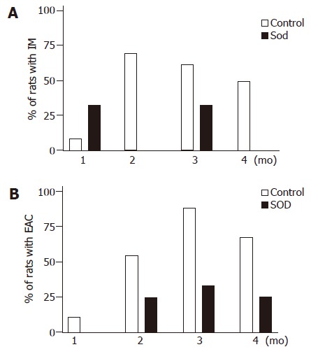
The exogenous administration of SOD did not significantly affect the development of inflammation, erosions, ulcers or metaplasia in continuity. However, SOD significantly reduced the risk of IM beyond the anastomotic level (odds ratio = 0.326, 95%CI = 0.108-0.981, P = 0.046) (Figure 6A). SOD also significantly decreased the risk of EAC (odds ratio = 0.243, 95%CI = 0.073-0.804, P = 0.021) (Figure 6B). In addition, esophageal mucosal levels of superoxide anion and SOD activity in rats with and without treatment were determined, confirming that administration of SOD significantly decreased superoxide anion production (53.7±10.11 cpm/mg vs 22±5.67 cpm/mg protein, P<0.01) and significantly increased SOD activity in rats that received treatment when compared to non-treated rats (28.2±4.72 U/g vs 13.4±1.82 U/g tissue, P<0.05).
Figure 6.

Incidence of intestinal metaplasia distant from the anastomosis site (A) and esophageal adenocarcinoma (B). The risk of both intestinal metaplasia and adenocarcinoma was significantly lower in the SOD-treated group than in the control group (P = 0.046 and P = 0.021, respectively) (n = 6-10).
DISCUSSION
Surgical models allowing gastro-duodeno-esophageal reflux or duodeno-esophageal reflux in rats are commonly used to produce experimental BE and EAC[14-17]. Most studies using these models analyzed the prevalence of BE and AC only at one point, but the sequence esophagitis-BE-dysplasia-AC has not been analyzed. The present study established the chronology of this sequence, which is especially useful for analyzing the effect of treatments. Thus, reactive changes and intestinal metaplasia in continuity to the anastomotic site appeared at high rates in the first month, while the prevalence of metaplasia distant from the anastomosis increased considerably in mo 2. High rates of AC were found in mo 4, so we considered this time of follow-up to be sufficient for the second part of the study, which evaluated the efficacy of exogenous administration of SOD in preventing different lesions. The next step of the study was to evaluate the involvement of free radicals, especially superoxide anion as a mechanism of damage in this model. Thus, we showed that the progression of normal mucosa to AC in the esophagus was associated with a parallel increase in superoxide anion mucosal levels. Superoxide anion remained increased until the end of the follow-up, suggesting that superoxide anion plays a pathogenic role in the development of EAC. Furthermore, in the present study we avoided administration of iron, since it has been shown to promote oxidative stress in similar models[18-20]. Therefore, our results provide evidence that gastroduodenal reflux per se induces oxidative stress and the development of EAC. Apart from superoxide anion, the involvement of other ROS in this model cannot be excluded. Superoxide anion is scavenged by SOD, generating hydrogen peroxide and oxygen. In this experimental study, a significant and progressive decrease of SOD activity was found, which was parallel to the increase of superoxide anion levels. When SOD activity was analyzed by different degrees of lesion, all groups showed a significantly lower SOD activity than control rats. These data suggest that the decrease of SOD activity may be in part responsible for the accumulation of superoxide anion observed and makes unlikely the generation of hydrogen peroxide in substantial amounts. Since nitric oxide is a natural scavenger of superoxide anion and overexpression of the inducible nitric oxide synthase has been reported in a similar model of EAC in rats[20], the search for generation of the ONOO– radicals in the mucosa is the next logical step. In the current study, substantial amounts of peroxynitrites analyzed by nitrotyrosine immunostaining were found in the esophagus of rats with esophagojejunostomy, suggesting that peroxynitrite formation is a common event in the presence of excess superoxide anion radicals in this model.
DNA damage resulting from exposure to ROS is believed to play a significant role in carcinogenesis. Though the spectrum of ROS-induced DNA lesions is quite extensive, the modified base 8-hydroxy-2’-deoxyguanosine (8-OHdG) is the most abundant and extensively studied because it can be easily measured[21]. In the present study, 8-OHdG levels were significantly increased at all the time points (1, 2, and 4 mo after esophagojejunostomy), though there were no differences between the different periods. These data indicate that oxidative DNA damage is a very early event and may explain why AC appears so fast in this model. Our results agree with previous studies showing an early increase of oxidative damage to DNA both in an EDA model in rats[22] and in the human esophagitis-metaplasia-dysplasia-AC sequence[23].
The progressive decrease of SOD activity may be due to two main causes: decrease in SOD expression and/or enzyme inactivation. Therefore, we investigated SOD expression by Western blot at different time points of follow-up. Blots revealed that Cu, Zn-SOD expression remained unchanged after esophagojejunostomy, while a progressive increase of MnSOD expression was found, which agrees with previous reports showing higher levels of MnSOD in esophageal carcinomas in comparison to normal tissue[24,25]. The observed increase in MnSOD expression may reflect a compensatory mechanism against the decrease of activity[11]. Therefore, enzyme inactivation is the more plausible mechanism underlying the low SOD activity found in this model. Since tyrosine nitration by peroxynitrite has been shown to inhibit the activity of MnSOD[11], we studied this possibility. We showed that MnSOD was partially tyrosine-nitrated in esophagi exposed to chronic reflux and the amount of nitrated MnSOD increased with time. The low level of nitrated MnSOD observed in some of the control esophagi may reflect the post-mortem ischemia or the basal tyrosine nitration levels under normal physiological conditions. Therefore, we suggest that nitration of MnSOD contributes to partially decrease the SOD activity, but other mechanisms known to inactivate SOD isoforms[26] such as oxidation are likely to be involved in this model.
Administration of various free-radical scavengers can prevent esophageal mucosal damage in different animal models of reflux esophagitis[5-8,27,28], where the most pronounced scavenging effect can be achieved with SOD. However, there is no information about the effect of SOD on the progression of esophageal mucosal damage to BE and EAC. In the present study, administration of SOD significantly reduced the risk of developing IM beyond the anastomotic level and also decreased the risk of EAC, being associated with a relative risk reduction of 68% and 76% respectively. The fact that SOD had no effect on inflammation is in discordance with previous studies demonstrating its effectiveness in preventing esophageal damage in experimental models of esophagitis. However, these studies evaluated the effect of SOD for a short period of time, just hours or at most a few days. Thus, it is possible that the scavenging effect of SOD might be sufficient to decrease mucosal damage in the short term but the magnitude of injury induced by continuous reflux extending over a much longer period of time overwhelms the ability of SOD to avoid mucosal inflammation and ulceration. However, the reduction of IM and AC achieved with the administration of SOD in this study suggests that SOD might be a therapeutic tool for preventing the development of BE and AC in patients with esophagitis. Though administration of SOD in such basis could not completely eliminate the generation of superoxide anion, treatment with SOD significantly lowered superoxide anion levels and increased SOD activity in rat esophagi, indicating that ontosein thus administrated can reach the esophageal mucosa and effectively scavenge superoxide anion at this level.
In conclusion, gastrointestinal reflux induces esophageal IM and AC in most animals undergoing esophagojejunostomy in mo 4. The development of these lesions is associated with esophageal superoxide anion and peroxynitrite generation, reduction of endogenous SOD activity due in part to MnSOD inactivation by nitration and early oxidative damage to DNA. Exogenous administration of SOD reduces the risk of IM and AC in this rat model of gastrointestinal reflux, which constitutes the first evidence showing that antioxidant treatment is effective in preventing BE and EAC.
ACKNOWLEDGMENTS
The authors thank Sara Serrano, Pilar Pina and Lidia Floría from the Service of Pathology for their invaluable technical assistance.
Footnotes
Supported by grants from CICYT (SAF2000-0123) and Instituto de Salud Carlos III (C03/02). Elena Piazuelo is supported by Instituto de Salud Carlos III and Instituto Aragonés de Ciencias de la Salud
Science Editor Wang XL and Guo SY Language Editor Elsevier HK
References
- 1.Blot WJ, Devesa SS, Fraumeni JF. Continuing climb in rates of esophageal adenocarcinoma: an update. JAMA. 1993;270:1320. [PubMed] [Google Scholar]
- 2.Olyaee M, Sontag S, Salman W, Schnell T, Mobarhan S, Eiznhamer D, Keshavarzian A. Mucosal reactive oxygen species production in oesophagitis and Barrett's oesophagus. Gut. 1995;37:168–173. doi: 10.1136/gut.37.2.168. [DOI] [PMC free article] [PubMed] [Google Scholar]
- 3.Wetscher GJ, Hinder RA, Klingler P, Gadenstätter M, Perdikis G, Hinder PR. Reflux esophagitis in humans is a free radical event. Dis Esophagus. 1997;10:29–32; discussion 33. doi: 10.1093/dote/10.1.29. [DOI] [PubMed] [Google Scholar]
- 4.Wetscher GJ, Hinder RA, Bagchi D, Hinder PR, Bagchi M, Perdikis G, McGinn T. Reflux esophagitis in humans is mediated by oxygen-derived free radicals. Am J Surg. 1995;170:552–556; discussion 552-556;. doi: 10.1016/s0002-9610(99)80014-2. [DOI] [PubMed] [Google Scholar]
- 5.Naya MJ, Pereboom D, Ortego J, Alda JO, Lanas A. Superoxide anions produced by inflammatory cells play an important part in the pathogenesis of acid and pepsin induced oesophagitis in rabbits. Gut. 1997;40:175–181. doi: 10.1136/gut.40.2.175. [DOI] [PMC free article] [PubMed] [Google Scholar]
- 6.Lanas A, Soteras F, Jimenez P, Fiteni I, Piazuelo E, Royo Y, Ortego J, Iñarrea P, Esteva F. Superoxide anion and nitric oxide in high-grade esophagitis induced by acid and pepsin in rabbits. Dig Dis Sci. 2001;46:2733–2743. doi: 10.1023/a:1012735714983. [DOI] [PubMed] [Google Scholar]
- 7.Wetscher GJ, Hinder PR, Bagchi D, Perdikis G, Redmond EJ, Glaser K, Adrian TE, Hinder RA. Free radical scavengers prevent reflux esophagitis in rats. Dig Dis Sci. 1995;40:1292–1296. doi: 10.1007/BF02065541. [DOI] [PubMed] [Google Scholar]
- 8.Wetscher GJ, Perdikis G, Kretchmar DH, Stinson RG, Bagchi D, Redmond EJ, Adrian TE, Hinder RA. Esophagitis in Sprague-Dawley rats is mediated by free radicals. Dig Dis Sci. 1995;40:1297–1305. doi: 10.1007/BF02065542. [DOI] [PubMed] [Google Scholar]
- 9.Valencia J, Velilla C, Urpegui A, Alvarez I, Llorens MA, Coronel P, Polo S, Bascón N, Escó R. The efficacy of orgotein in the treatment of acute toxicity due to radiotherapy on head and neck tumors. Tumori. 2002;88:385–389. doi: 10.1177/030089160208800507. [DOI] [PubMed] [Google Scholar]
- 10.Soteras F, Lanas A, Fiteni I, Royo Y, Jimenez P, Iñarrea P, Ortego J, Esteva F. Nitric oxide and superoxide anion in low-grade esophagitis induced by acid and pepsin in rabbits. Dig Dis Sci. 2000;45:1802–1809. doi: 10.1023/a:1005521925785. [DOI] [PubMed] [Google Scholar]
- 11.MacMillan-Crow LA, Crow JP, Kerby JD, Beckman JS, Thompson JA. Nitration and inactivation of manganese superoxide dismutase in chronic rejection of human renal allografts. Proc Natl Acad Sci USA. 1996;93:11853–11858. doi: 10.1073/pnas.93.21.11853. [DOI] [PMC free article] [PubMed] [Google Scholar]
- 12.Pittman KM, MacMillan-Crow LA, Peters BP, Allen JB. Nitration of manganese superoxide dismutase during ocular inflammation. Exp Eye Res. 2002;74:463–471. doi: 10.1006/exer.2002.1141. [DOI] [PubMed] [Google Scholar]
- 13.Huang X, Powell J, Mooney LA, Li C, Frenkel K. Importance of complete DNA digestion in minimizing variability of 8-oxo-dG analyses. Free Radic Biol Med. 2001;31:1341–1351. doi: 10.1016/s0891-5849(01)00681-5. [DOI] [PubMed] [Google Scholar]
- 14.Fein M, Ireland AP, Ritter MP, Peters JH, Hagen JA, Bremner CG, DeMeester TR. Duodenogastric reflux potentiates the injurious effects of gastroesophageal reflux. J Gastrointest Surg. 1997;1:27–32; discussion 33. doi: 10.1007/s11605-006-0006-x. [DOI] [PubMed] [Google Scholar]
- 15.Fein M, Peters JH, Chandrasoma P, Ireland AP, Oberg S, Ritter MP, Bremner CG, Hagen JA, DeMeester TR. Duodenoesophageal reflux induces esophageal adenocarcinoma without exogenous carcinogen. J Gastrointest Surg. 1998;2:260–268. doi: 10.1016/s1091-255x(98)80021-8. [DOI] [PubMed] [Google Scholar]
- 16.Miwa K, Sahara H, Segawa M, Kinami S, Sato T, Miyazaki I, Hattori T. Reflux of duodenal or gastro-duodenal contents induces esophageal carcinoma in rats. Int J Cancer. 1996;67:269–274. doi: 10.1002/(SICI)1097-0215(19960717)67:2<269::AID-IJC19>3.0.CO;2-6. [DOI] [PubMed] [Google Scholar]
- 17.Seto Y, Kobori O. Role of reflux oesophagitis and acid in the development of columnar epithelium in the rat oesophagus. Br J Surg. 1993;80:467–470. doi: 10.1002/bjs.1800800420. [DOI] [PubMed] [Google Scholar]
- 18.Chen X, Yang Gy, Ding WY, Bondoc F, Curtis SK, Yang CS. An esophagogastroduodenal anastomosis model for esophageal adenocarcinogenesis in rats and enhancement by iron overload. Carcinogenesis. 1999;20:1801–1808. doi: 10.1093/carcin/20.9.1801. [DOI] [PubMed] [Google Scholar]
- 19.Goldstein SR, Yang GY, Curtis SK, Reuhl KR, Liu BC, Mirvish SS, Newmark HL, Yang CS. Development of esophageal metaplasia and adenocarcinoma in a rat surgical model without the use of a carcinogen. Carcinogenesis. 1997;18:2265–2270. doi: 10.1093/carcin/18.11.2265. [DOI] [PubMed] [Google Scholar]
- 20.Goldstein SR, Yang GY, Chen X, Curtis SK, Yang CS. Studies of iron deposits, inducible nitric oxide synthase and nitrotyrosine in a rat model for esophageal adenocarcinoma. Carcinogenesis. 1998;19:1445–1449. doi: 10.1093/carcin/19.8.1445. [DOI] [PubMed] [Google Scholar]
- 21.Marnett LJ. Oxyradicals and DNA damage. Carcinogenesis. 2000;21:361–370. doi: 10.1093/carcin/21.3.361. [DOI] [PubMed] [Google Scholar]
- 22.Chen X, Ding YW, Yang Gy, Bondoc F, Lee MJ, Yang CS. Oxidative damage in an esophageal adenocarcinoma model with rats. Carcinogenesis. 2000;21:257–263. doi: 10.1093/carcin/21.2.257. [DOI] [PubMed] [Google Scholar]
- 23.Sihvo EI, Salminen JT, Rantanen TK, Rämö OJ, Ahotupa M, Färkkilä M, Auvinen MI, Salo JA. Oxidative stress has a role in malignant transformation in Barrett's oesophagus. Int J Cancer. 2002;102:551–555. doi: 10.1002/ijc.10755. [DOI] [PubMed] [Google Scholar]
- 24.Janssen AM, Bosman CB, van Duijn W, Oostendorp-van de Ruit MM, Kubben FJ, Griffioen G, Lamers CB, van Krieken JH, van de Velde CJ, Verspaget HW. Superoxide dismutases in gastric and esophageal cancer and the prognostic impact in gastric cancer. Clin Cancer Res. 2000;6:3183–3192. [PubMed] [Google Scholar]
- 25.Izutani R, Asano S, Imano M, Kuroda D, Kato M, Ohyanagi H. Expression of manganese superoxide dismutase in esophageal and gastric cancers. J Gastroenterol. 1998;33:816–822. doi: 10.1007/s005350050181. [DOI] [PubMed] [Google Scholar]
- 26.MacMillan-Crow LA, Crow JP, Thompson JA. Peroxynitrite-mediated inactivation of manganese superoxide dismutase involves nitration and oxidation of critical tyrosine residues. Biochemistry. 1998;37:1613–1622. doi: 10.1021/bi971894b. [DOI] [PubMed] [Google Scholar]
- 27.Lee JS, Oh TY, Ahn BO, Cho H, Kim WB, Kim YB, Surh YJ, Kim HJ, Hahm KB. Involvement of oxidative stress in experimentally induced reflux esophagitis and Barrett's esophagus: clue for the chemoprevention of esophageal carcinoma by antioxidants. Mutat Res. 2001;480-481:189–200. doi: 10.1016/s0027-5107(01)00199-3. [DOI] [PubMed] [Google Scholar]
- 28.Oh TY, Lee JS, Ahn BO, Cho H, Kim WB, Kim YB, Surh YJ, Cho SW, Hahm KB. Oxidative damages are critical in pathogenesis of reflux esophagitis: implication of antioxidants in its treatment. Free Radic Biol Med. 2001;30:905–915. doi: 10.1016/s0891-5849(01)00472-5. [DOI] [PubMed] [Google Scholar]


