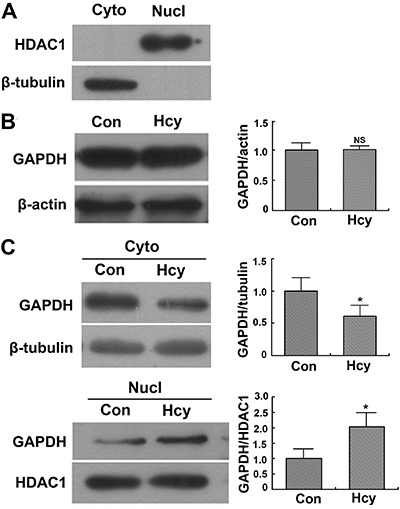
Figure 4. Translocation of glyceraldehyde 3-phosphate dehydrogenase (GAPDH) detected by Western blotting. Cytoplasmic (Cyto) and nucleic (Nucl) proteins were separated from Neuro2a cells incubated in the presence of 5 mM homocysteine (Hcy) or absence (Con) of Hcy for 48 h. The protein levels of GAPDH in subcellular fractions were detected by Western blotting with beta-tubulin as control. A, Purification of subcellular fractions was confirmed with the nucleic marker protein histone deacetylase 1 (HDAC1) and cytoplasmic protein marker beta-tubulin. B, Total GAPDH protein level. C, GAPDH protein levels in cytoplasm and nucleus. *P<0.05 (Student-Newman-Keuls test). NS: not significant.

