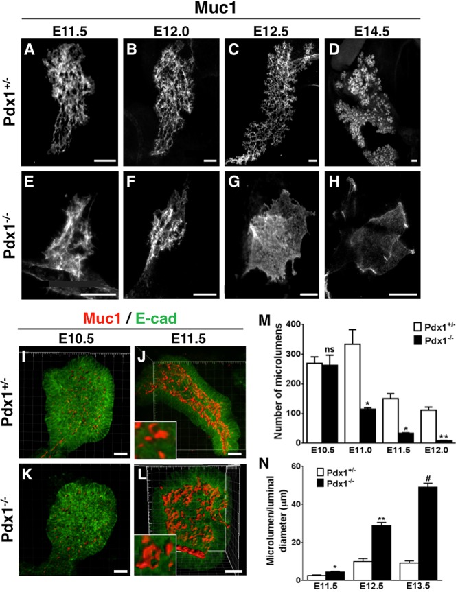
Fig. 1.
In the absence of Pdx1, the pancreatic ductal plexus transforms into a bag-like cyst. (A-H) Z-stack images of whole-mount tissues stained with Muc1 to visualize lumens, reconstructed using 3D projection (ImageJ). (I-L) Imaris surface reconstruction of lumens (Muc1, red; E-cad, green). Note that whereas E10.5 microlumen size is similar in Pdx1+/− and Pdx1−/− buds (I,K), lumen sizes become expanded in Pdx1−/− by E11.5 (L) when compared with Pdx1+/− (J). Representative microlumens included for comparison (insets, panels J,L). (M) Quantification of number of microlumens at different stages. (N) Comparison of microlumen diameter as lumens start to fuse. ns, not statistically significant, *P<0.05, **P<0.01, #P<0.001 (Student's t-test). Scale bars: 50 μm in A-H; 25 μm in I-L.

