Fig. 6.
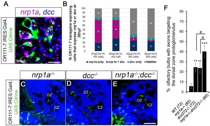
Relative contributions of Nrp1a and Dcc to DZ mistargeting. (A) A single optical section through a 36 hpf or111-7 transgenic embryo (frontal view). Cell bodies are in green. Dorsal is up and medial is to the right. nrp1a mRNA is in magenta and dcc mRNA is in blue. (B) The percentages of or111-7-labeled neurons expressing nrp1a, dcc or both at 27, 36, 48 or 60 hpf. (A,B) nrp1a and dcc are co-expressed in a large portion of or111-7-labeled neurons during early development (asterisks in A). (C-E) Single optical sections through 72 hpf or111-7 transgenic larvae (frontal view). Axons are in green. Dorsal is up and medial is to the right. Propidium iodide (blue) labels cell bodies revealing protoglomeruli as cell-free regions. (F) The percentage of OBs with projections to the DZ protoglomerulus. (C-F) nrp1a−/− mutants, dcc−/− mutants and nrp1a−/−;dcc−/− double mutants have an increase in projections to the DZ as compared with wild-type siblings. nrp1a−/−;dcc−/− double mutants have a statistically significant increase in DZ errors when compared with single-mutant siblings. Statistical significance was estimated using two-tailed (**P<0.01, ***P<0.001) or one-tailed (#P<0.05) Fisher's exact tests. Error bars represent s.e. of the sample proportion. CZ, central zone; DZ, dorsal zone. Scale bars: 10 µm in A; 25 µm in C-E.

