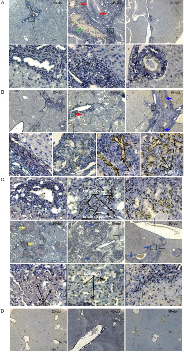
Fig. 2.

2. Immunohistochemistry of iNOS in the liver of mice infected with C. sinensis. Liver sections were incubated with the antibody against iNOS and visualized by DAB as brown-colored deposits, and hematoxylin was used to counterstain the nuclei in blue. The pathological changes in the livers of mice infected with different numbers of metacercariae, namely, 20 (A), 40 (B), and 80 (C), were detected at 20, 45, and 90 days post-infection (dpi). Normal uninfected mice served as controls (D). The original magnification was at 40×, and the upper/lower corresponding images were magnified at 400×. Arrows in red, blue, yellow, and green highlight the ductular adenomatous hyperplasia, fibrous tissue hyperplasia, spotty necrosis of hepatic lobules, and fluke, respectively. The figure shows 1 representative biopsy out of 6. SECs, sinusoidal endothelial cells; LDCs, liver dendritic cells; KCs, Kupffer cells; BECs, biliary epithelial cells. +, iNOS positive; -, iNOS negative.
