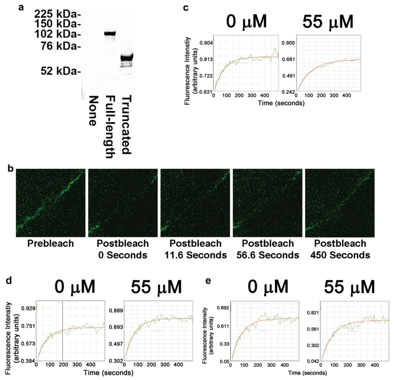
Fig. 5.
H2O2 treatment slows the movement of full-length GFP-occludin into the tight junction region but not movement of GFP-occludin within the apical membrane or C-terminal truncated GFP-occludin into the tight junction region. (a) Western blot analysis of expression of GFP-tagged proteins in untransduced MDCK cells (None), MDCK cells transduced with full-length GFP-occludin construct (Full-length) or MDCK cells transduced with C-terminal truncated GFP-occludin construct (Truncated). FRAP analysis was performed on MDCK cell populations transduced with full-length GFP-occludin construct and maintained in the absence or presence of 55 μM H2O2 for 2 h. GFP-occludin in an area of the tight junction region of transduced MDCK cells was photobleached. (b) Images of this region were obtained prior to, immediately following and at increasing amounts of time following photobleaching. The images shown are obtained from transduced MDCK cells maintained in the absence of H2O2. (c) The fluorescence signal in the photobleached area was quantitated and plotted as a function of time for transduced MDCK cells treated with 0 μM H2O2 or 55 μM H2O2. (d) Representative traces for FRAP analysis of full-length GFP-occludin moving within the apical membrane as a function of time for transduced MDCK cells treated with 0 μM or 55 μM H2O2. (e) Representative traces for FRAP analysis of C-terminal truncated GFP-occludin moving into the tight junction region as a function of time for transduced MDCK cells treated with 0 μM or 55 μM H2O2.

