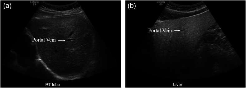
Fig. 1.
US of (a) normal versus (b) fatty liver. The texture of the right image is in general grainier, but some speckling, inherently caused by the US medium, can be seen on the left image as well. The portal vein shows up more clearly in the left image as fatty liver obscures the vessel interface. Additionally, fatty liver is more hyperechoic. Compared to the evenly bright normal liver, the top of the right US image (near the surface) is much brighter than the bottom (deeper) as more sound waves are immediately reflected in the fatty liver.

