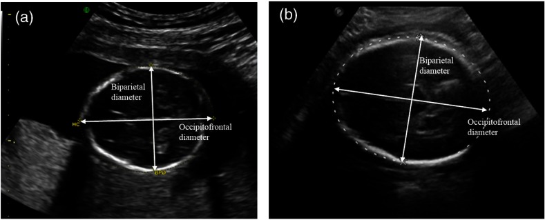
Fig. 3.
US of a fetus (a) who will develop craniosynostosis and (b) one who will not. By manual inspection, the difference between the two is not obvious at this stage. This is further complicated by the imaging medium, where the front and back of the skull are sometimes indistinct, as shown in the right image.

