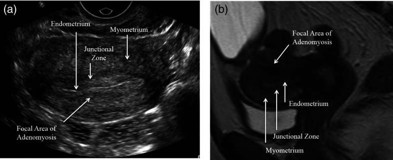
Fig. 5.
(a) US and (b) a sagittal MRI through the uterus showing adenomyosis from the same patient. The uterine lining is much clearer and more easily distinguished from the background in the MRI and the shape of the uterus more easily assessed. In the MRI, a focal region of dark signal resulting in abnormal thickening of the junctional zone is clearly seen. By US, the region of focal adenomyosis is difficult to identify from that of surrounding normal myometrium.

