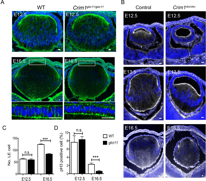
Fig. 5.
Crim1glcr11 and Crim1cko mice exhibit lens development defects. (A,B) F-actin staining with Phalloidin in Crim1glcr11 (A) and in Crim1cko (B) lenses reveals a small lens of altered morphology. Images shown are representative of six independent experiments. (C,D) Quantification of total LE cell number (C) and the percentage of LE cells undergoing proliferation (D) at E12.5 and E16.5. n=4; ***P<0.001 (Student's t-test). Scale bars: 20 μm.

