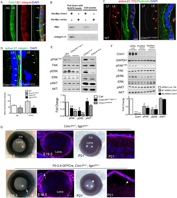
Fig. 6.
Crim1 regulates integrin and FAK and ERK phosphorylation in lens development. (A) Colocalization of β1 integrin and Crim1 (arrows) at LE cell-cell adhesions and at the basal surface of LE cells. (B) Co-immunoprecipitation of β1 integrin with His-Myc-tagged Crim1-FL in HEK 293T cells. (C,D) Immunostaining of active (9EG7) β1 integrin shows decreased staining in Crim1glcr11 (C) and Crim1null (D) lenses. Arrows point to the LE cell basement membrane. Phalloidin stains the actin cytoskeleton; DAPI stains nuclei. Quantification of fluorescence intensity is shown beneath. n=3. (E) Western blot analysis of P6 wild-type and Crim1glcr11 lenses (left) and E18.5 control and Crim1null lenses (right) with the indicated antibodies. n=5. (F) 21EM15 cells were treated with either of two siRNAs directed against Crim1 for 48 h and then cell lysates were western blotted with the indicated antibodies. Each bar represents the mean of triplicates. (G) Compound heterozygous P0-3.9-GFPCre;Crim1flox/+;Itgb1flox/+ mice exhibit iris coloboma (arrows) and abnormal lens morphology at E18.5, and later develop bilateral cataract at P21 (arrowheads). n=6; all six P21 compound heterozygotes obtained were affected, as compared with none of four littermate controls. Arrowhead in P21 section indicates LE cell detachment from LF cells. Red, Phalloidin-stained actin cytoskeleton. Blue, DAPI-stained nuclei. *P<0.05, **P<0.01, ***P<0.001 (Student's t-test). Scale bars: 10 μm.

