
Fig. 1.
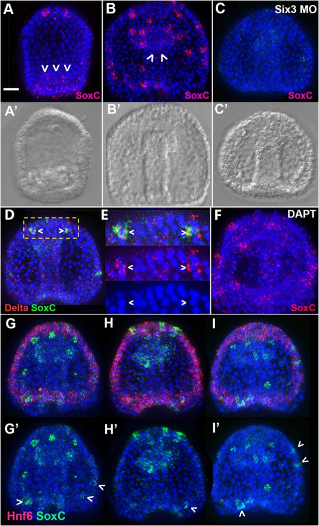
SoxC expression in control sea urchin embryos and embryos treated with DAPT or Six3 morpholino. (A,B) In situ hybridization for SoxC (red) shows signal in individual cells of the anterior end (apical plate) and posterior end (vegetal plate; arrowheads) of a 28 h embryo (A) and in lateral ectoderm and foregut (arrowheads) of a 48 h embryo (B). (C) SoxC expression in all regions is greatly reduced in Six3 morphants. (A′,B′,C′) The corresponding DIC images. (D,E) SoxC (green) transcripts are detected in some cells expressing Delta (red, arrowheads). Doubly labeled cells are shown at higher magnification in E. (F) SoxC-expressing cells (red) form small clusters in DAPT-treated embryos. (G-I) Three embryos illustrating that the locations and number of SoxC-expressing cells (green) vary among embryos. Magenta signal is the ciliated band/APD marker Hnf6. Arrowheads indicate SoxC-expressing cells in the ciliated band. (G′,H′,I′) The corresponding green channels. All images are ventral (oral face) views with the animal pole at top. All embryos except A are at the 2-day late gastrula stage. Nuclei are labeled with DAPI (blue). Scale bar: 20 µm.