
Fig. 3.
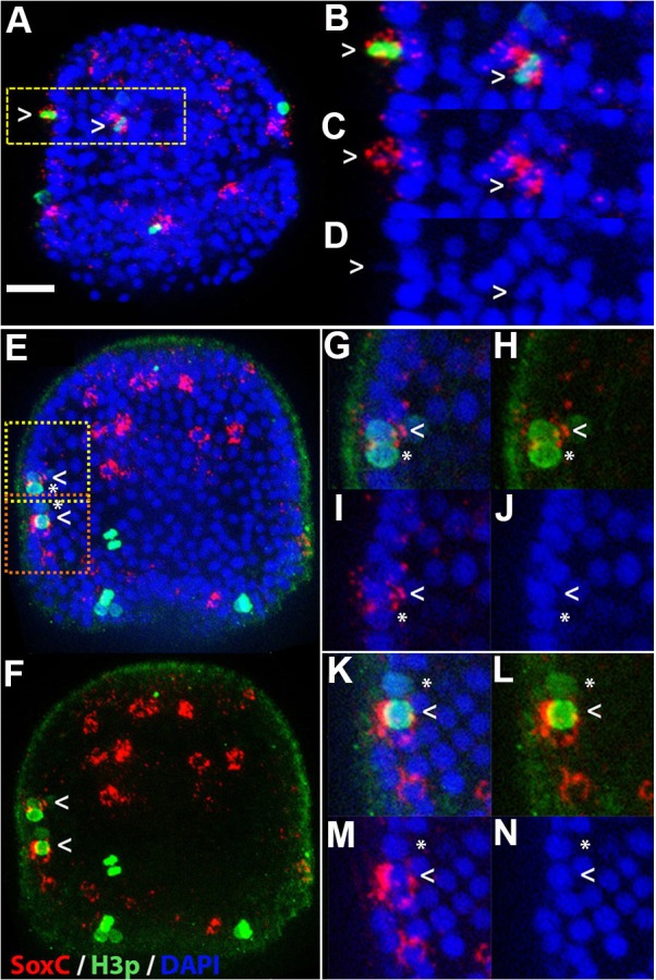
Some dividing cells express SoxC. Embryos were immunostained with the H3p antibody, which labels cells from prophase to anaphase, and hybridized with a probe for SoxC mRNA. (A) Two SoxC-expressing cells (red) are H3p positive (green, arrowheads). (B) Higher magnification image of H3p-positive cells outlined by the yellow box in A. (C) SoxC (red) and DAPI (blue). (D) DAPI only. (E) Two adjacent cells: one SoxC positive (arrowhead) and the other SoxC negative (asterisk). (F) The SoxC and H3p channels of E. (G-J) Higher magnification images of the region in E outlined in yellow. (K-N) Higher magnification images of the region in E outlined in brown. All embryos are at 66 h and are shown with anterior poles at the top. Scale bar: 20 µm.