
Fig. 5.
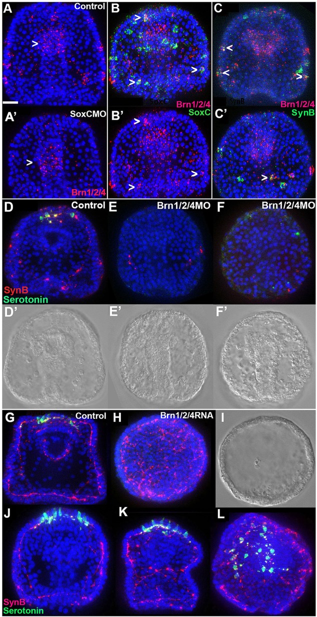
Brn1/2/4 is required downstream of SoxC for differentiation of all neurons. (A,A′) Brn1/2/4 (red) is expressed in individual cells in ectoderm and in a group of contiguous cells in the foregut (arrowhead) of a control embryo (A); the number of individual labeled cells is greatly reduced in ectoderm, whereas expression in contiguous endoderm cells (arrowhead) is not detectably affected in SoxC morphants (A′). (B,B′) Brn1/2/4 and SoxC doubly labeled in situ hybridization of an embryo, showing a few Brn1/2/4-expressing cells (red) that also express SoxC (green, arrowheads). (B′) Red channel only. (C,C′) Doubly labeled in situ hybridization for Brn1/2/4 and SynB in two different embryos, showing that some Brn1/2/4-expressing cells (red) also express SynB (green, arrowheads). (D-F) Brn1/2/4 morpholino greatly reduced the number of neurons (green, serotonin; red, SynB). (D) Control; (E,F) two morphants. (D′-F′) Corresponding DIC images. (G-I) Immunostaining for serotonin (green) and SynB (red) in a 3-day control embryo (G) and a Brn1/2/4 mRNA-injected (H) embryo (higher dose). The latter embryo is an epithelial sphere that lacks serotonin staining but has expanded SynB signal. (I) DIC image of embryo in H. (J-L) Three different injected embryos showing that, at the lower Brn1/2/4 mRNA dose, the serotonergic cell number is increased. At ∼1.0 µg/µl Brn1/2/4 mRNA, most embryos resembled that shown in H; at 0.5 µg/µl, more embryos resembled those in J-L. Embryos in A-C are at 46 h and in D-L at 72 h. All embryos are shown with the anterior poles at the top, except H,I,L; L is an anterior view of the APD. Nuclei are labeled with DAPI (blue). Scale bar: 20 µm.