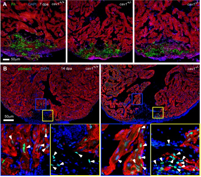
Fig. 7.
Fibronectin deposition and Tgfβ signaling activation in cav1 mutants. (A) Section images of injured ventricles from the three cav1 genotypes at 7 dpa. Sections are stained for Tnnt (red) and fibronectin (Fn, green). (B) Sections of injured ventricles at 14 dpa are stained for Tnnt (red) and pSmad3 (green). The boxed regions are enlarged to show details. Arrowheads indicate pSmad3-positive CMs (blue box, Tnnt+) and non-CMs (yellow box, Tnnt−).

