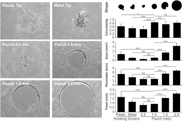
Figure 1. Morphometric analysis of epithelial wounds.
Stratified cultures of human corneal epithelial cells were injured by rotation using a plastic or metal tip (rotating scratch model), or using dermal punchs (0.5, 1.0, 1.5 or 2.0 mm diameter) followed by epithelial debridement (punch model). Morphology of the injured area was analyzed by measuring main parameters of the wound shape, including circularity, area, perimeter and Feret diameter. The area and perimeter of the wounds ranged from 0.3 to 3.3 mm2 and 2.9 to 7.0 mm, respectively. The values for circularity and Feret diameter ranged from 0.48 to 0.87 and 0.8 to 2.1 mm, respectively. Use of plastic or metal tips produced modest results in terms of shape and circularity. Punch injuries followed by epithelial debridement using a disposable plastic tip (i.e., 1.0, 1.5 and 2.0 mm punch) produced consistent shapes and high circularity values. Results are displayed as mean +/− SD (N = 9). Significance was determined using one-way ANOVA with Bonferroni’s post-hoc test. *p < 0.05; ***p < 0.001. ns, non-significant.

