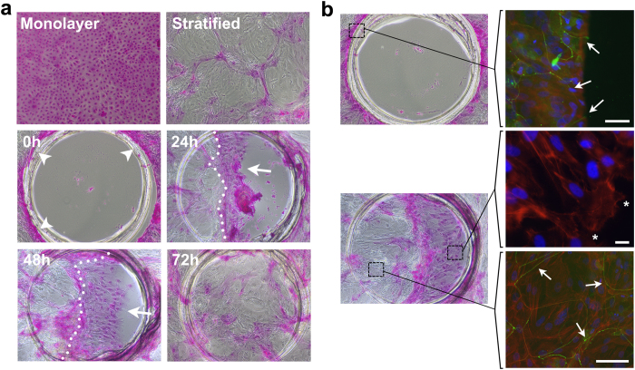
Figure 5. Barrier function is restored as epithelial cells migrate en bloc.
(a) Circular wounds of 1 mm in diameter were made using a punch template. The extent of rose bengal dye penetrance was assessed at 0, 24, 48 and 72 h following culture in serum-containing medium. Induction of stratification in control cultures resulted in the formation of islands of stratified cells that excluded the dye. A defined staining of the wound margins was observed immediately after wounding (arrowheads). Epithelial migration was characterized by uptake of dye by cells at the leading edge (arrows) but not behind the stratification front (dotted line). Closure of the wound at 72 h was associated with the complete restoration of barrier function. (b) For immunofluorescence microcopy, stratified cultures in chambered slides were wounded using a 1.5 mm punch and manually scraped using a pipette tip. Occludin (green) was detected at the cellular margin after wounding (arrows), and was restored to the intercellular junctions within the stratification front during migration. Cells at the leading edge (asterisks) extended lamellipodial protrusions as shown by actin staining (red) and were characterized by lack of occludin localization at the cell-cell junction. Nuclei were counterstained using DAPI (blue). Scale bars, 50 μm (top), 20 μm (middle), 100 μm (bottom).

