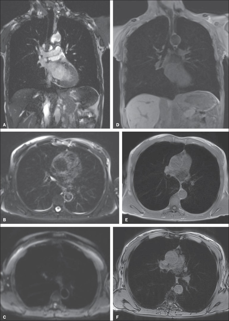
Figure 6.
(A-F). Image examples for the suggested protocol. A: Coronal T2-weighted FSE TrueFisp sequence (4 mm). B: Axial, T2-weighted sequence Blade axial (4 mm). C: Axial, diffusion-weighted image (5 mm). D: Coronal, T1-weighted sequence VIBE with fat suppression (4 mm). E,F: T1-weighted sequence in- and out-of-phase.
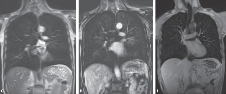
(G–I): Image examples for the suggested protocol. G: Coronal, T2-weighted HASTE sequence. H: Coronal, T1-weighted perfusion sequence. I: T1-weighted sequence VIBE with fat suppression.


