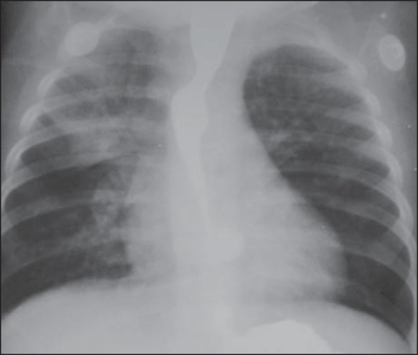
Figure 4.

A seven-month-old male child with repetition pneumonia. Anteroposterior view of the chest with esophageal contrast-enhancement. Opacity is observed in the right upper lobe, compatible with pneumonia. Concentric narrowing of the lumen of the proximal esophageal third, with upstream dilatation. Such findings are strongly suggestive of extrinsic compression by double aortic arch. After surgical correction, the respiratory symptoms and the esophageal compression disappeared.
