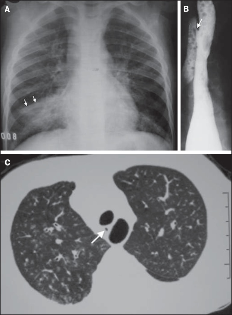
Figure 5.
A six-year-old boy with repetition pneumonia. A: Chest radiography, anteroposterior view showing subtle diffuse opacities in both lungs, confluent in the middle lobe. The caudal sift of the horizontal scissure (arrows) characterizes the presence of atelectatic component. B: Esophagography demonstrating H-type fistula to the trachea (arrow). C: High resolution computed tomography, axial section at the level of the fistula characterized by the dark dot (arrow) between the esophagus (with air) and the trachea. Centrilobular opacities, some of them branching, demonstrating involvement of small airways.

