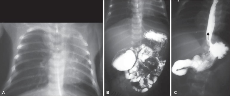
Figure 7.
A neonate with Down syndrome and respiratory symptoms. A: Anteroposterior chest radiography showing left lung with decreased volume and transparency. Fading of the left cardiac silhouette indicates upper lobe atelectasis. B: EGDS. Small bowel transit shows partial obstruction at the level of the second duodenal portion, with appearance suggestive of duodenal diaphragm (windsock sign). C: Secondary gastroesophageal reflux.

