
Figure 1.
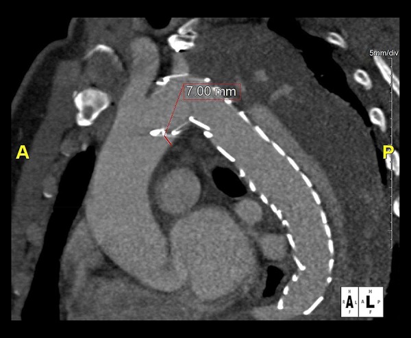
Displayed is a parasagittal multiplanar reformation of the postprocedural CT of the patient with the type I endoleak. After previous left subclavian artery revascularization the patient had undergone TEVAR for chronic aortic dissection Stanford type B (patient #19; Tables 2 and 3). The conformability analysis showed a 7 mm distance between the gold band of the endograft and the inner curvature of a type III aortic arch.