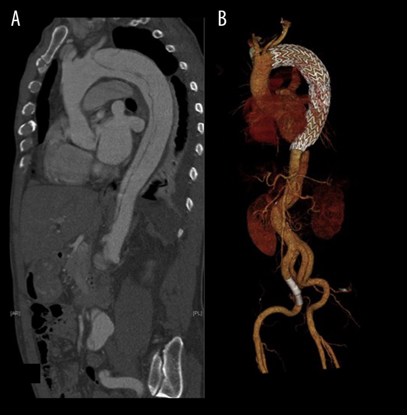
Figure 2.

Displayed are a preoperative sagittal reconstruction (A) of a CT-angiography in 61-year-old patient, who was treated for acute complicated aortic dissection Stanford type B (patient #15; Tables 2 and 3) and a 3D-reconstruction (B) of a follow-up CT scan in the same patient, showing favorable conformability of the endograft within the aortic arch.
