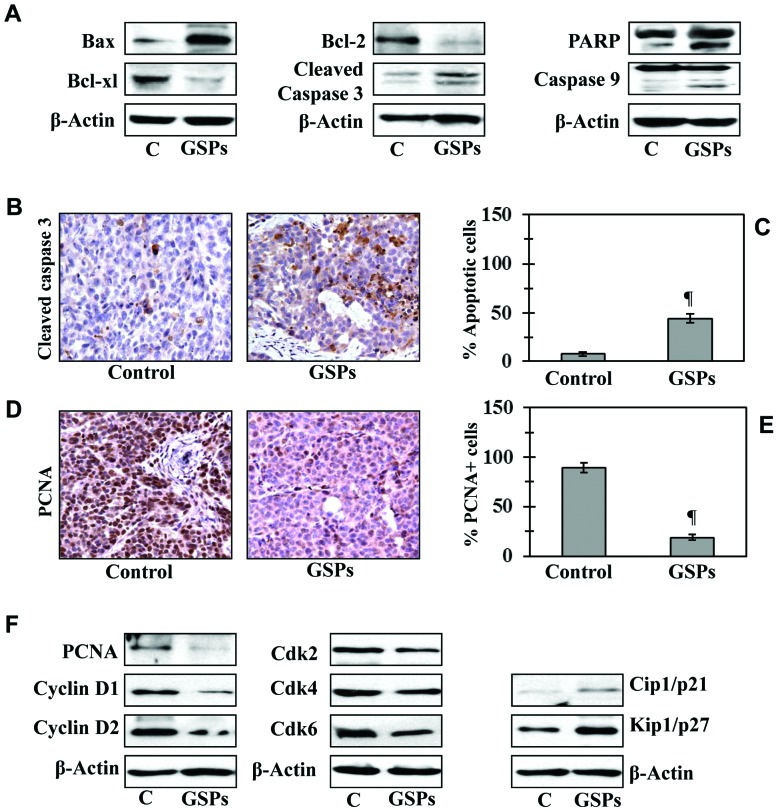
Figure 4.
Dietary GSPs induce apoptosis and inhibit tumor cell proliferation in tumor xenograft tissues. Experiment was terminated at 40 days post inoculation of A375 cells, xenograft tissues were harvested for the analysis of biochemical markers. (A) Western blot analysis of proteins of apoptotic family in tumor lysates of control mice and mice that were fed on diet supplemented with 0.5% GSPs. (B) Representative photomicrographs showing immunohistochemical detection of cleaved caspase-3+ cells in xenograft sections. (C) Number of cleaved caspase-3+ cells (apoptotic cells) were counted under a microscope at 3–4 different places of a section. (D) Representative photomicrographs are presented showing the PCNA+ cells in different treatment groups. PCNA+ cells are shown in dark brown. (C and E) Immunohistochemical staining data are presented as mean percentage of positive cells ± SD, n=3/group. Significant difference vs. non-GSPs-treated control group; ¶P<0.001. (F) Western blot analysis of the proteins of cell cycle regulation, PCNA and tumor suppressors in tumor lysates of control and GSPs-treated groups.

