Figure 4.
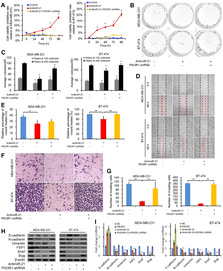
AntimiR-21-induced suppression of proliferation, clonogenicity, invasiveness, and metastatic properties of breast cancer cells is mediated by direct repression of PIK3R1. (A) MTS assays were conducted on breast cancer cells after transfection with antimiR-21 (50 nmol/l), antimiR-21 + PIK3R1-shRNA or control. At 48, 72 and 96 h, the antimiR-21 lines showed significantly reduced levels of proliferation as compared to control lines. PIK3R1-shRNA reversed the effect of antimiR-21 on cells. (B) Representative images depicting clonogenic assays performed with cells plated at 200 cells/well. (C) In MDA-MB-231 and BT-474 lines, antimiR-21 resulted in a decrease in colony number as compared to control lines. PIK3R1-shRNA reversed the effect of antimiR-21 on cells. (D) Representative images depicting cell migration assays. (E) Cell migration was quantitated as percentage of wound-healed area from corresponding control and transfected cells. (F) Invasion assays in these control and transfected cells. (G) For each cell line, antimiR-21 resulted in reduced invasion as compared to controls. PIK3R1 knockdown reversed the effect of antimiR-21 on cell migration in both cell lines. (H) MDA-MB-231 and BT-474 lines were transfected with PIK3R1, antimiR-21, antimiR-21 + PIK3R1-shRNA or control, followed by western blot analysis of the indicated EMT-related proteins. Relative E-cadherin, N-cadherin, vimentin, FSP1, snail and slug levels were normalized to the β-actin level. (I) Breast cancer lines were transfected with PIK3R1, antimiR-21, antimiR-21 + PIK3R1-shRNA or control, followed by RT-qPCR analysis of the indicated EMT-related mRNAs. Data represent mean ± SD. *P<0.05; **P<0.01.

