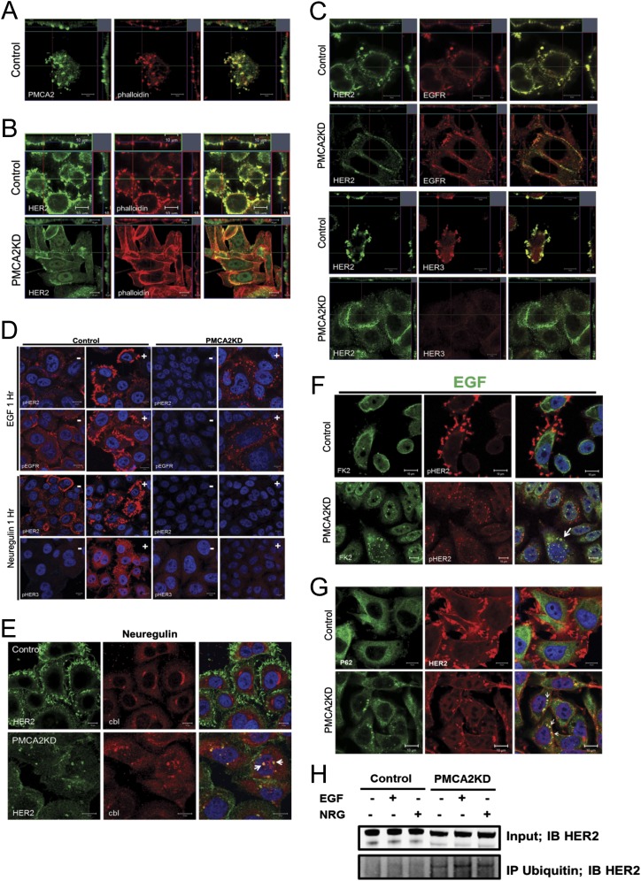
Fig. S3.
(A) Immunofluorescence confocal images for costaining for PMCA2 (green) and actin (phalloidin, red) in SKBR3 cells. (Right) Merged images. (Insets) Z-stacks of the cells in two different orientations. Note that PMCA2 and actin colocalize within apical membrane protrusions. (B) Immunofluorescence confocal images for costaining for HER2 (green) and actin (phalloidin, red) in control and PMCA2KD SKBR3 cells. (Right) Merged images. (Insets) Z-stacks of the cells in two different orientations. In control cells, HER2 is localized within actin-rich membrane domains protruding from the apical surface of the cell. However, knocking down PMCA2 leads to reorganization of the actin cytoskeleton, flattening of the cells, and more diffuse membrane staining for HER2. (C) Immunofluorescence confocal images of costaining for HER2 (green) and EGFR (red) in the top two rows and HER2 (green) and HER3 (red) in the bottom two rows. (Right) Merged images. (Insets) Z-stacks as before. Note colocalization of HER2 with EGFR or HER3 in membrane protrusions in control SKBR3 cells (rows 1 and 3). However, in PMCA2 KD cells, staining for HER2 and EGFR becomes more diffusely distributed and less well colocalized in the membrane (row 2). Furthermore, in PMCA2 KD cells, HER3 staining is much reduced and no longer colocalizes with HER2 (row 4). (D) Staining for pHER2 or pEGFR in serum-free media (−) or after 1 h treatment with EGF (+) (top two rows). In control cells, treatment with EGF leads to prominent localization of pHER2 and pEGFR within membrane protrusions. However, in PMCA2KD cells, treatment with EGF leads to internalization of both pHER2 and pEGFR. Bottom two rows: Staining for pHER2 or pHER3 in serum-free media (−) or after 1 h treatment with neuregulin (+). In control cells, treatment with neuregulin leads to prominent localization of pHER2 within membrane protrusions, although pHER3 becomes mostly cytoplasmic within 1 h of stimulation. In PMCA2KD cells, treatment with neuregulin causes almost no stimulation of pHER3. (E) Costaining for HER2 and c-Cbl in control (Top) and PMCA2KD (Bottom) cells after 2 h treatment with neuregulin. Arrows point to intracellular colocalization of HER2 with c-Cbl only in knock-down cells. (F) Costaining for pHER2 with FK2 in control (Top) and PMCA2KD (Bottom) cells after 2 h treatment with EGF. (Right) Merged images. Arrows point to intracellular colocalization of pHER2 with FK2 in knockdown cells after stimulation with EGF. (G) Costaining for HER2 with p62 in control (Top) and PMCA2KD (Bottom) cells after 2 h treatment with EGF. (Right) Merged images. Arrows point to intracellular colocalization of HER2 with p62 in knockdown cells treated with EGF. (H) Co-IP of polyubiquitin complexes with HER2 in control and PMCA2KD cells in serum-free media or after 2 h of treatment with EGF or NRG. HER2 is pulled down with antibody against polyubiquitin only in the knock-down cells. (Scale bars, 10 μm.)

