Fig. 1.
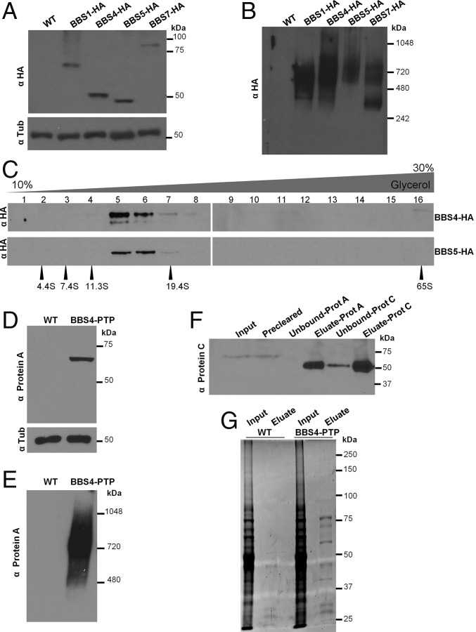
TbBBS proteins assemble into the TbBBSome. (A) Western blot for 3× HA epitope-tagged TbBBS proteins. Wild type (WT) was used as negative control. Tubulin (Tub) blot served as loading control. (B) Western blot for HA-tagged TbBBS proteins by blue native PAGE. (C) Western blot for TbBBS4-HA and TbBBS5-HA in 10–30% (vol/vol) glycerol gradient centrifugation. Numbers 1–16 indicate gradient fractions. Protein standards of known S values are shown at the bottom. (D) Western blot for PTP epitope-tagged TbBBS4. WT was used as negative control. Tub blot served as loading control. (E) Western blot for TbBBS4-PTP by blue native PAGE. (F) Western blot for tandem purification of TbBBS4-PTP probed with anti-Protein C. TEV protease was used for elution before Protein C purification, hence the reduction in size. (G) SYPRO Ruby protein stain for tandem purification of TbBBS4-PTP. Eluates were used for shotgun proteomics (SI Appendix, Tables S1 and S5).

