Fig. 2.
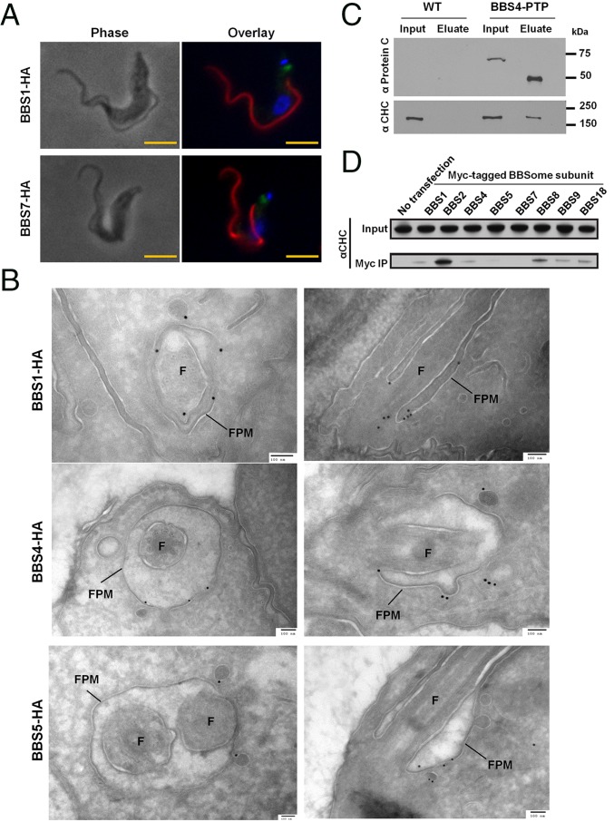
The TbBBSome localizes to the flagellar pocket membrane and interacts with clathrin. (A) IF for TbBBS-HA proteins: TbBBS-HA (green), PFR2 (red), and DAPI (blue). (Scale bar: 5 µm.) (B) Immunogold labeling for TbBBS-HA proteins. Cross-sections and longitudinal sections of the flagellar pocket are shown in Left and Right, respectively. Flagellar pocket membrane (FPM) and flagellum (F) are indicated. (Scale bar: 100 nm.) Immunogold quantification is provided in SI Appendix, Table S2. (C) Western blot of TbBBS4-PTP immunoprecipitation by Protein A probed with anti-Protein C and anti-CHC. TEV protease was used to elute TbBBS4-PTP, hence the reduced size of TbBBS4 in the eluate. Input (1×) and eluate (8×) in anti-Protein C blot; input (1×) and eluate (100×) in anti-CHC blot. (D) Western blot for CHC in immunoprecipitation (IP) of Myc-tagged BBSome proteins from HEK293 cells.

