Fig. 4.
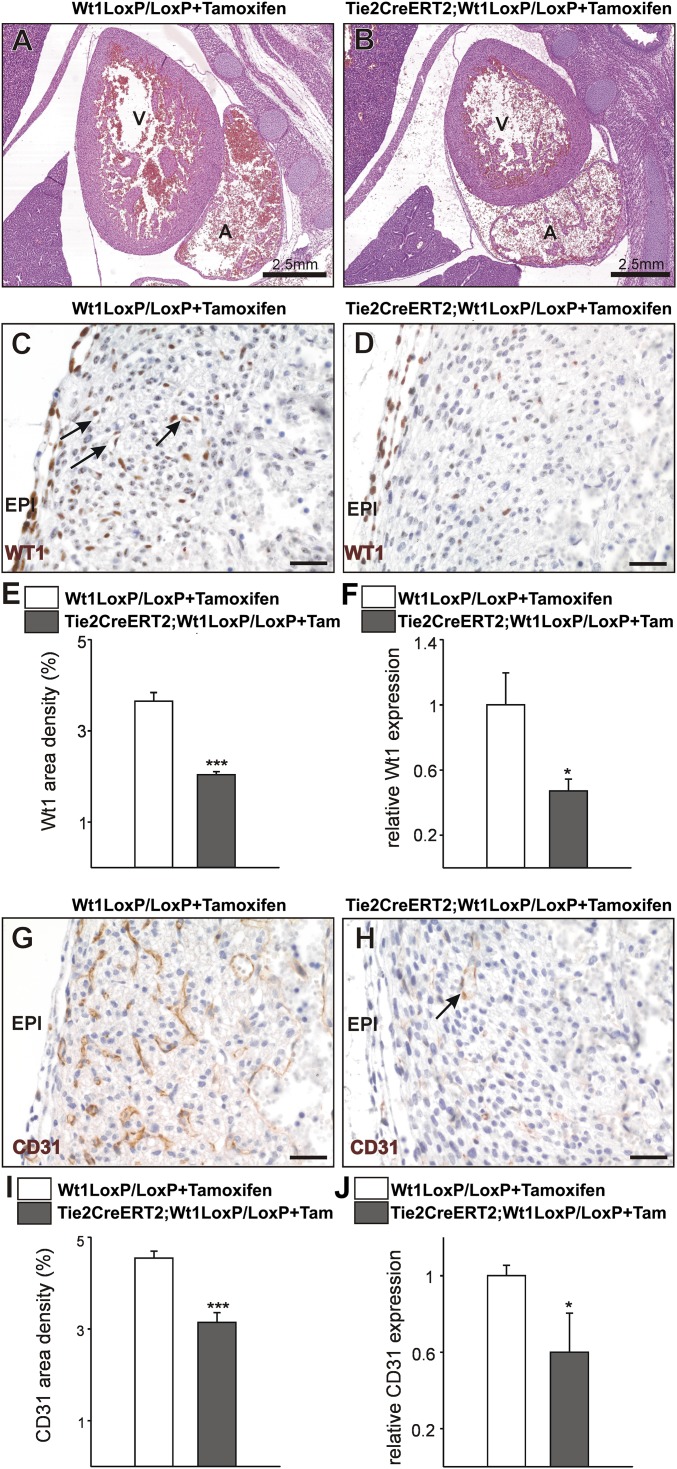
Endothelial Wt1 expression is required for coronary vessel formation. (A and B) H&E-stained sections from control Wt1LoxP/LoxP+Tamoxifen (A) and mutant TieCreERT2;Wt1LoxP/LoxP+Tamoxifen (B) E16.5 embryos. (C–F) The WT1+ cell contribution to coronary vessels (arrows in C) is reduced in the mutants (D and E), and Wt1 gene expression in E16.5 mutant hearts is reduced (F). (G and H) E16.5 mutant embryonic hearts show a decrease in compact ventricular wall coronary CD31+ cells (arrow in H; compare with G). (I and J) Quantification of the area occupied by CD31+ cells (I) and CD31 gene expression (J) in E16.5 hearts. A, atrium; EPI, epicardium; V, ventricle. (Scale bars: 50 µm.) Data are mean ± SEM; *P < 0.05, ***P < 0.001.

