Fig. S3.
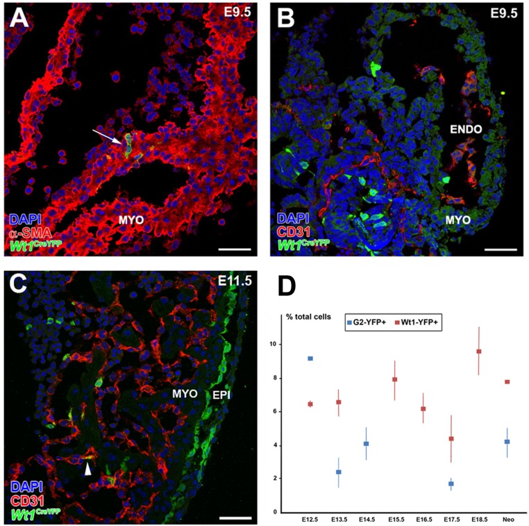
Wt1 lineage-derived cardiomyocytes in the E9.0 mouse heart. (A and B) A minority of Wt1CreYFP+ cells can be found in the developing heart before or during PE cell transference to the myocardial surface (E9.5). These Wt1CreYFP+ cells express myocardial markers (α-SMA; arrow in A) but not vascular markers (CD31+) (B). (C) At early epicardial stages (E10.5–11.5) some rare endocardial Wt1CreYFP+/CD31+ cells could be recorded (arrowhead). (D) The total percentage of cardiac Wt1CreYFP+ and G2CreYFP+ cells throughout the course of development is shown. ENDO, endocardium; EPI, epicardium; MYO, myocardium. (Scale bars: 50 µm.)

