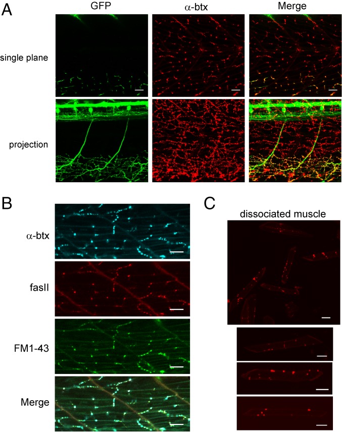
Fig. 1.
En passant neuromuscular synapses between the CaP motor neuron and target fast muscle. (A) Sample images of two segments of an SAIGFF213A/UAS:GFP transgenic fish with labeled CaP motor neurons and α-btx–labeled acetylcholine receptors. A single image plane is shown in Upper, and a projection through the fish is shown in Lower. The merge is shown in Right. (B) Distribution of α-btx, fasII, and FM1-43 labels in ventral muscle. (C) Dissociated muscle cells labeled with α-btx. Upper shows a field with multiple muscle cells, and three examples of individual cells are shown in Lower. (Scale bars: 20 μm.)

