Fig. 2.
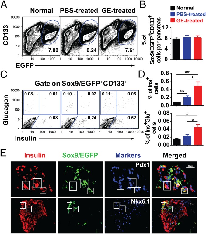
Long-term administration of low-dose GE increases Insulinlo cells among Sox9/EGFP+CD133+ cells, although it does not increase the percentage of Sox9/EGFP+CD133+ in diabetic mice with medium hyperglycemia. After treated by TM and Alloxan as described in Fig. S2A, the pancreatic tissues from normal and diabetic Sox9CreERT2 R26mT/mG mice with medium hyperglycemia after 56-d treatment with PBS or GE were dissociated into single cells. Sox9/EGFP+CD133+, Insulin+, and Insulin+Glucagon+ cell populations were analyzed by flow cytometry. (A and B) The percentage of Sox9/EGFP+CD133+ among total pancreatic mononuclear cells is shown (mean ± SEM, n = 4). (C and D) After gating on Sox9/EGFP+CD133+ cell population, the percentage of Insulin+ and Insulin+Glucagon+ cells is shown. (E) Pancreatic tissues were stained for Insulin, EGFP, and Pdx1 or NKX6.1. One representative staining pattern of islets in mice is shown from 4 replicate experiments. Triple-positive cells are shown in the box. (Scale bars: 30 µm.) *P < 0.05; **P < 0.01; ***P < 0.001.

