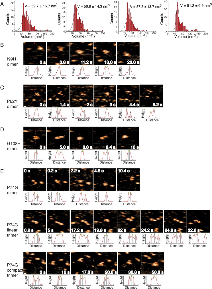
Fig. S6.
Mutations to SOD1 trimer interfaces stabilize formation of nonnative extended dimer conformations. (A) Volume distributions of (left to right) I99H, P74G, P62Y, and G108H after 24 h of aggregation at 30 μM SOD1 at 37 °C indicate dominance of a nonnative dimer species. Solid lines are Gaussian fits, with most probable volume designated. Errors are ±SD. (B) I99H-SOD1, a trimer-destabilizing mutant, exists almost exclusively as an extended dimer, as demonstrated by HS-AFM images. (C) P62Y-SOD1, a trimer-destabilizing mutant, exists as an extended dimer, as demonstrated by HS-AFM images. (D) G108H-SOD1, a trimer-destabilizing mutant, exists as an extended dimer with a small trimeric population (not shown), as demonstrated by HS-AFM images. (E) P74G-SOD1, a trimer-stabilizing mutant, exists as an extended dimer and linear or compact trimer. In B–E, the scan area is 50 nm × 50 nm. (Scale bar: B–E, 5 nm.) Dashed lines on the image represent the 1D cross-section used for height profiling below each image, with red circles indicating the initial point of measurement. Green dots on the height profiles indicate peak positions, with the distance between the two gray lines representing the distance of two adjacent subunits. Data were obtained with regular AFM imaging of dried samples.

