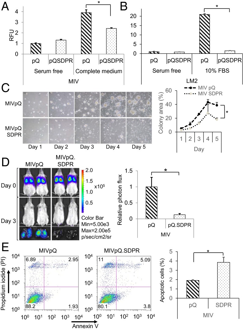
Fig. 4.
SDPR primes MIV cells for apoptosis and inhibits extravasation. (A) Transendothelial cell migration potential of the control and MIVpQ.SDPR cells toward serum-free or complete media was assessed 48 h after the seeding by calcein staining, P = 0.0374. RFU, relative florescence unit. (B) Effect of SDPR on the extravasation potential of LM2 cells was quantified, P = 7.87479E-07. (C) Growth of control and MIVpQ.SDPR cells were monitored over time in 3D cell culture and quantified, on the Right, by measuring colony area, P = 0.01. (D) Effect of SDPR overexpression on survivability of MIV cells was monitored 72 h after the tail vein injections by Caliper IVIS Spectrum. Whole-animal imaging is presented on the Upper Left, and extracted lungs are shown on Lower Left. Quantification of cell survivability was assessed on the Right, based on photon flux, P = 0.0014, npQ = 5, npQSDPR = 8. (E) Annexin V and PI staining were used to assess the basal level of apoptosis in control and MIVpQ.SDPR cells. Quantification of three independent Annexin V experiments is shown, P = 0.04. *P < 0.05.

