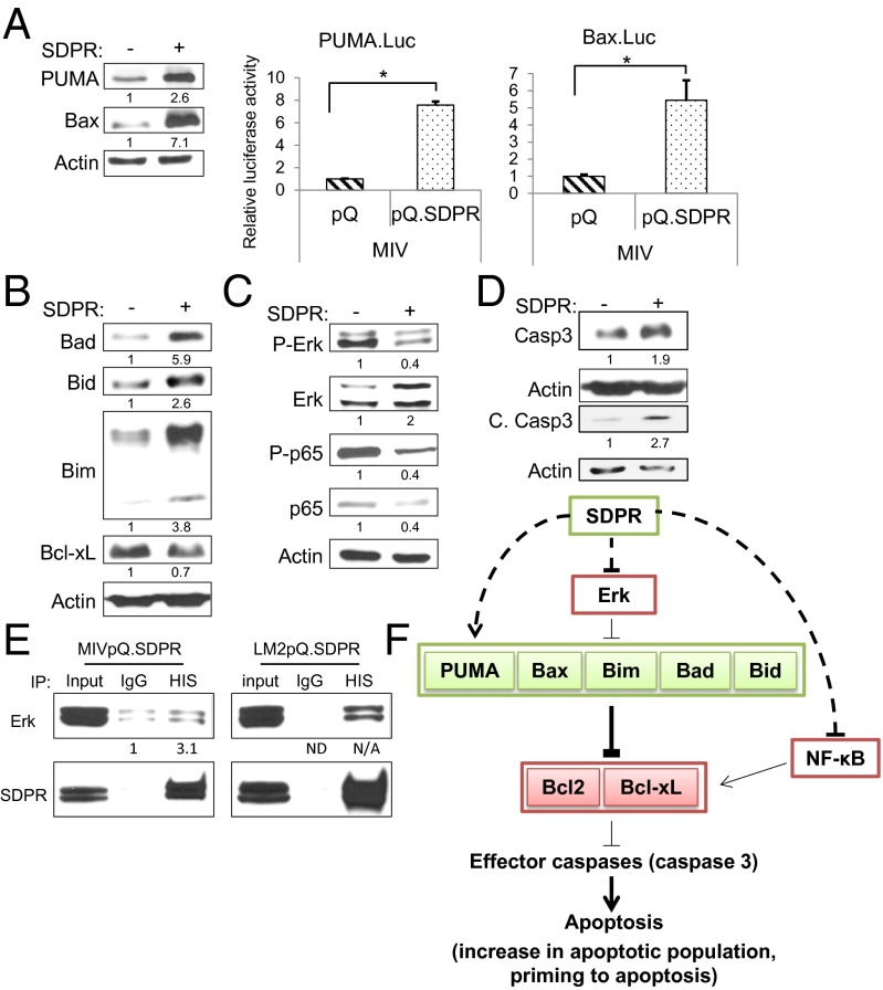
Fig. 5.
SDPR function and apoptosis. (A) The effect of SDPR overexpression on the proapoptotic PUMA and Bax expression in MIV cells was evaluated by Western blotting and luciferase reporter assays, pPUMA = 0.000003, pBax = 0.03 for all n = 3. (B) Protein levels of proapoptotic Bcl2 family members, Bad, Bid, and Bim, and antiapoptotic Bcl-xL were measured by Western blotting in control and MIpQ.SDPR cells. (C) The effect of SDPR overexpression on the activity of ERK and NF-κB pathways was evaluated by Western blotting against phosphorylated Erk and p65 protein levels, respectively. (D) Total and cleaved caspase-3 protein levels in control and MIVpQ.SDPR cells were measured by Western blotting. (E) Co-IP was carried out in MIV and LM2 cells using HIS (mouse) antibody to precipitate SDPR and mouse IgG as control. Western blot was used to assess the levels of precipitated SDPR and coprecipitated Erk. (F) SDPR overexpression, directly or indirectly, causes increases in proapoptotic BCL-2 family proteins. Additionally, the levels of the antiapoptotic protein Bcl-xL and the activity of prosurvival Erk and NF-κB signaling pathways were decreased due to SDPR overexpression. Overall, SDPR can alter the balance of regulatory proteins in the apoptotic pathway to favor cell death. *P < 0.05.

