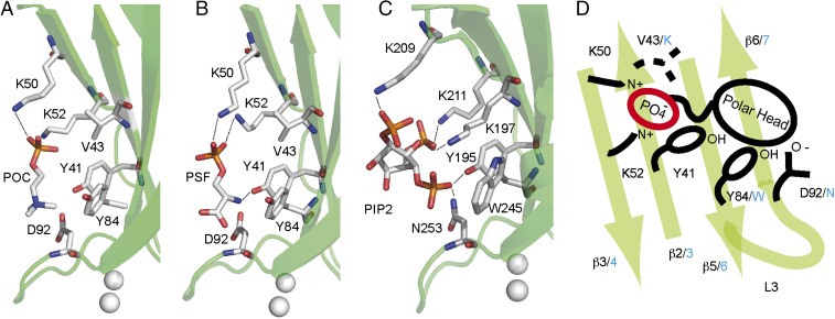
Fig. 2.
The polybasic lipid binding site of CAR4. (A) Structure of CAR4 in complex with POC and (B) with PSF. (C) Structure of the C2 domain of PKC-α in complex with PI(4,5)P2 is shown for comparison. The C2 domains are represented as green ribbons. Residues involved in ligand binding are represented as sticks. (D) Schematic representation of the polybasic lipid binding site highlighting the similarities and differences between CAR4 and PKC-α. Key CAR4 amino acids are labeled in black, and the corresponding amino acids in PKC-α are indicated in blue.

