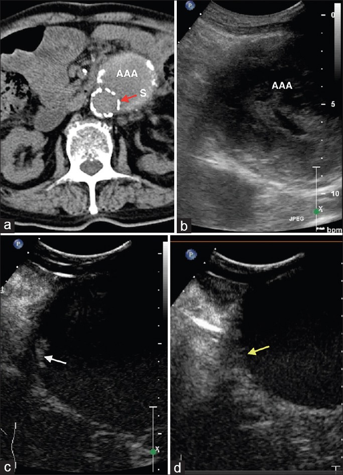
Figure 1.

A 84-year-old male patient, postendovascular aneurysm repair. (a) Computed tomography angiography shows no endoleak. S: Stent (red arrow). (b) Gray-scale ultrasound. (c) Contrast-enhanced ultrasound shows a small endoleak lateral to the upper end of the stent (type Ia) (white arrow). (d) After re-intervention, contrast-enhanced ultrasound shows no endoleak (yellow arrow).
